SPECT-CT SPINE GALLERY

Click on any image to enlarge

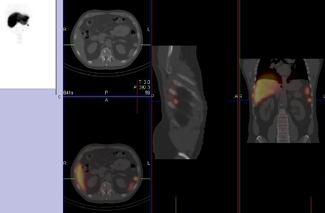
Transverse section perfusion lung scan with SPECTCT

Coronal section perfusion lung scan with SPECTCT

Sagittal section perfusion lung scan with SPECTCT
On all the lung perfusion images there is greater perfusion posteriorly as the patient’s are scanned lying down and the posterior alveoli are effectively compressed together and have higher aeration and perfusion per cubic centimetre.

An example of myocardial perfusion SPECT scan showing an abnormal area of perfusion in the lateral wall. The Bull’s eye on the left represent perfusion of the myocardium after stress, the middle image at rest, and the right image shows the difference. The hatched area is considered ischaemia and represents inadequate coronary reserve.

Sestamibi uptake in the left ventricle on a SPECT-CT scan.
This liver spleen SPECT-CT scan shows accessory splenic tissue. This excluded the lesion as a possible tumour seen on a prior CT scan of the abdomen.
The image below show a 3D surface rendered SPECT-CT of a sacral fracture with the colour representing isotope uptake in the fracture.


