Connectivity with Iain Duncan
xSPECT changed the diagnosis 20% of the time

The clinical value of xSPECT bone versus SPECT in bone scanning. Iain Duncan 2017*

xSPECT bone v SPECT bone in hybrid imaging A head to head comparison of 200 cases* ” template=”/nas/content/live/driainduncan/wp-content/plugins/nextgen-gallery/products/photocrati_nextgen/modules/ngglegacy/view/gallery-caption.php” order_by=”filename” order_direction=”ASC” returns=”included” maximum_entity_count=”500″]   xSPECT bone is a new reconstruction algorithm** which integrates information from the CT scan with the raw data from the bone SPECT acquisition to produce a higher resolution SPECT image, known as […]

Back pain and Bone scans (updated 2017)

Bone scans of the spine have become a clinically useful part of the evaluation of back pain. Uptake seen on a SPECT-CT correlates with metabolic activity in bone and the increased resolution and specificity of SPECT-CT has allowed a greater number and variety of clinically relevant findings. Most patients with back pain will have an […]

What is xSPECT bone?

A mobile friendly slideshow of this post can be found here. I have noted elsewhere about the dramatic transformation between “the old” and “the new” nuclear medicine that has taken place over the last decade (see my post Nuclear Medicine = New Clear Medicine). The fusion between nuclear medicine and CT was given the description SPECT-CT  -the SPECT […]

Nuclear Imaging in Prosthetic Joints

NUCLEAR TRACERS used in imaging prosthetic joints

The tracers discussed in this article are:

Tc-MDP: bone scintigraphy

Tc-sulesomab:  in vivo labelled Fab fragment of IgG1.  While this also binds to neutrophils (5%) and therefore should mimic WBC labelled scans it has a non-specific accumulation at infected sites. 35% of activity at 24hrs is in the bone marrow.

WBC labelled scans: in vitro labelled patient WBC with either Tc or In-111

Gallium 67

FDG (Fludeoxyglucose 18F): is also used for infection imaging but PET scanning is not discussed in this article.


SUMMARY OF OUR APPROACH TO SCANNING PROSTHESIS

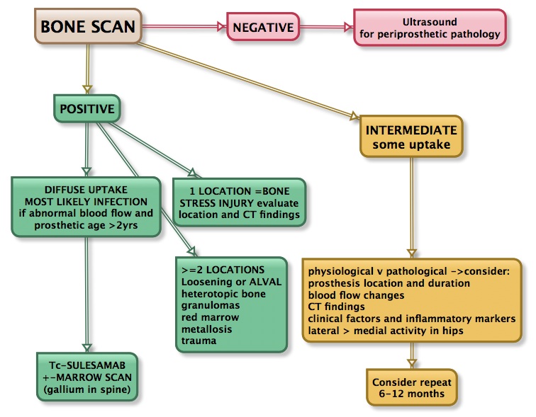
PROSTHETIC JOINT WORKFLOW

WORKFLOW for prosthetic joint complications


[…]

STRESS FRACTURES

STRESS FRACTURE GRADING GRADE XRAY SPECT-CT BONE SCAN MRI 1 normal mild /diffuse increase in tracer uptake. CT normal Increased periosteal T2 signal. Normal marrow signal. 2 normal moderately intense tracer uptake, mild sclerosis or thickening, but no fracture line plus changes on STIR and T2 sequences 3 Visible line intense (linear) focal tracer uptake with definite […]

Nuclear Imaging in Prosthetic Joints

NUCLEAR TRACERS used in imaging prosthetic joints The tracers discussed in this article are: Tc-MDP: bone scintigraphy Tc-sulesomab (trade name LeukoScan):  in vivo labelled Fab fragment of IgG1.  While this also binds to neutrophils (5%) and therefore should mimic WBC labelled scans it has a non-specific accumulation at infected sites. 35% of activity at 24hrs is […]

Perfusion Scintigraphy versus 256-slice CT Angiography in Pregnant Patients Suspected of Pulmonary Embolism: Comparison of Radiation Risks

Perisinakis K, Seimenis I, Tzedakis A, Damilakis J J Nucl Med. 2014;55:1273-1280 Summary The goal of this anthropomorphic phantom-based simulation investigation was to compare the dose exposure estimates of state-of-the-art 256-slice CT pulmonary angiography (CTPA) with lung perfusion scintigraphy (LPS) in pregnant patients with suspected pulmonary embolism (PE) at different body sizes and gestation times (the end of […]