
A mobile friendly slideshow of this post can be found here.
I have noted elsewhere about the dramatic transformation between “the old” and “the new” nuclear medicine that has taken place over the last decade (see my post Nuclear Medicine = New Clear Medicine). The fusion between nuclear medicine and CT was given the description SPECT-CT -the SPECT meaning “single photon emission computerised tomography” and representing cross sectional nuclear medicine. The CT represents the much better know computerised tomography or “CAT” scan. The bottleneck of SPECT-CT remained the limited resolution of the SPECT component of the fused imaging. The limitation seemed fundamental as a gamma wave cannot provide the spatial resolution of an x-ray. Siemens however came up with an ingenious solution by using the detailed spatial information about the tissues from the CT scan to reinterpret the gamma wave (SPECT) data.
In their publications Siemens notes “Using CT-based segmentation in the reconstruction, xSPECT Bone achieves a sharper definition of bony margins and lesion boundaries. The resulting reconstructed SPECT images (xSPECT Bone) appear to truly reflect the distribution of metabolically active bone within the skeletal system with sharper differentiation between cortical and spongy bone in the vertebrae, as well as in flat bones like the pelvis, scapula and skull. This also provides fine detail which helps differentiate between the cortex and marrow cavity in long bones and improved separation of bone from joint spaces—both for large joints such as the knee and shoulder and for small bones such as carpals and tarsal bones.”
The dramatic difference in the SPECT data can be appreciated even by the amateur. Fig 1 shows the xSPECT data on a single slice of the human pelvis and Fig 2 the exact same slice using conventional SPECT bone reconstruction (images courtesy of Garran Medical Imaging).
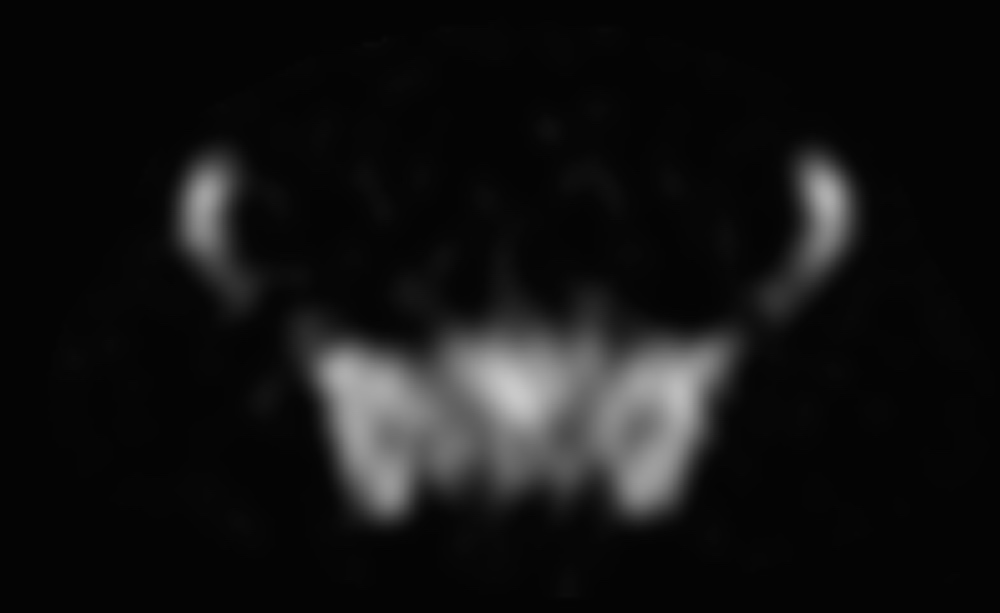
Fig 2. Single SPECT slice through the pelvis
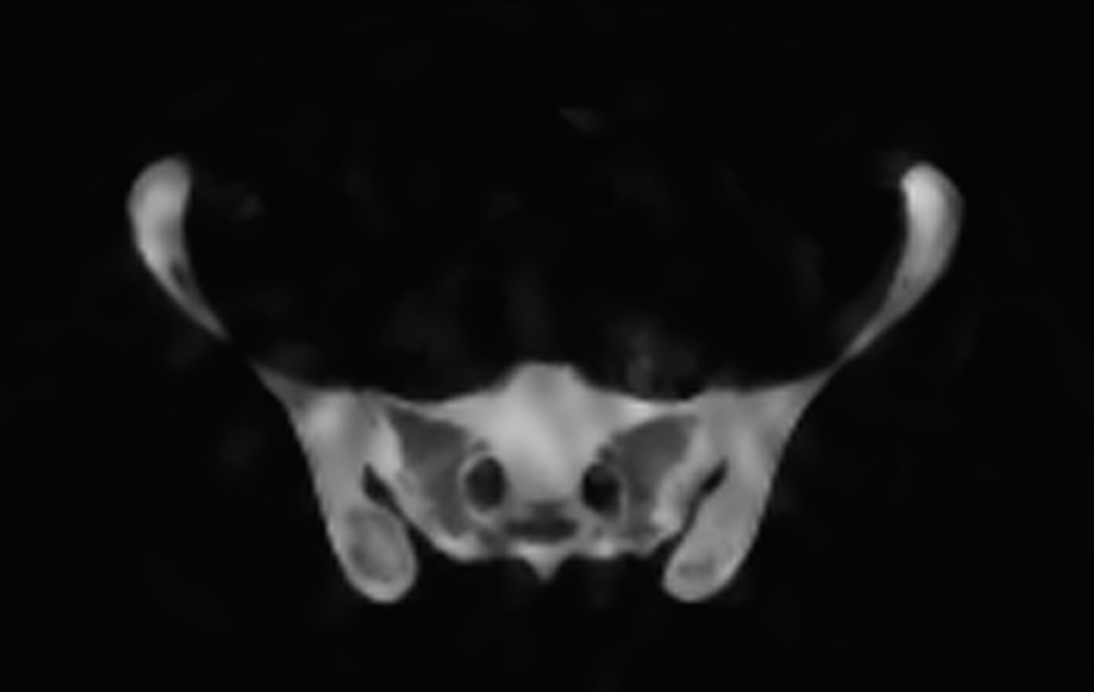
Fig 1. Single xSPECT slice through the pelvis
This advance has side stepped the physical limitation of gamma-ray imaging by using x-ray data to “re-engineer” the raw data obtained from the gamma camera and redistribute the uptake according to the know properties of each tissue voxel obtained direct from the CT (x-ray) data. The bone uptake resolution is taken to a new level thus bypassing the previous bottleneck.
What does this mean clinically? Well it is early days but the cases below all show bone isotope uptake in some very small locations that were previously rarely identified on SPECT-CT (again images courtesy Garran Medical Imaging).
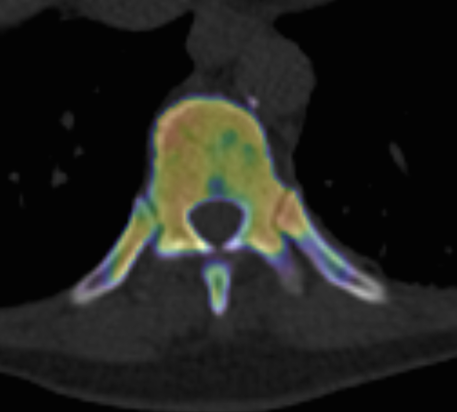
Fig3. Costovertebral joint arthrosis
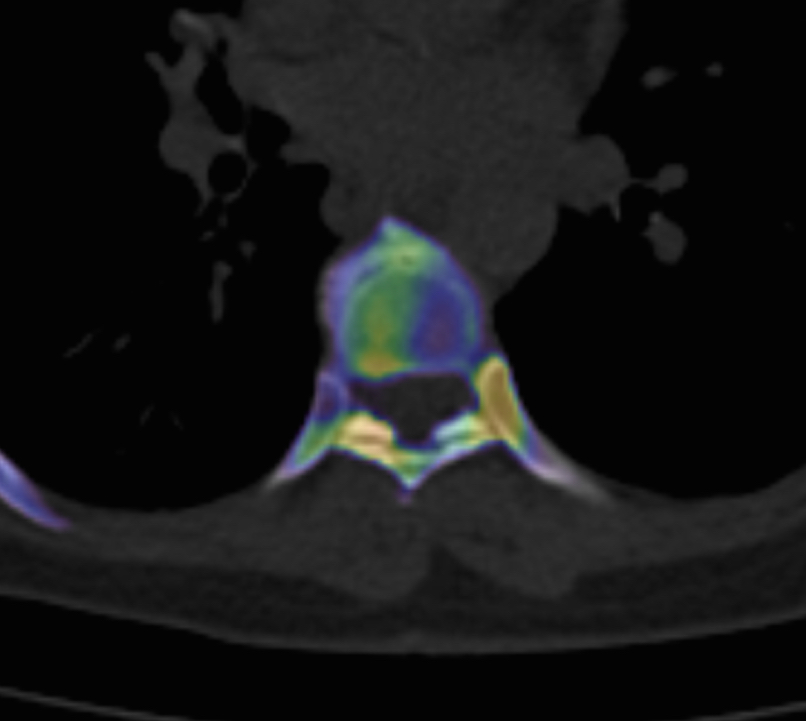
Fig4. Costotransverse and opposite thoracic facet joint arthrosis

Fig5. Thoracic facet joint arthrosis xSPECT-CT
All of these xSPECT-CT scans (Figs 3-5) show abnormalities that could not be localised with conventional SPECT-CT alone. How frequent these abnormalities are, how clinically important they are, and how often patient management is impacted are questions not yet answered.
Needless to say I am excited about the prospects that this technology will make a real difference to many patients and doctors, and I hope to release our early clinical data on this website soon. My experiences were presented at ANZSNM 2016 in Rotorua, New Zealand.
For nuclear medicine professionals a lecture on xSPECT from RSNA 2014 given by Professor Jerry Froelich can be found here.