
xSPECT bone v SPECT bone in hybrid imaging
A head to head comparison of 200 cases*


xSPECT bone is a new reconstruction algorithm** which integrates information from the CT scan with the raw data from the bone SPECT acquisition to produce a higher resolution SPECT image, known as xSPECT. The clinical value of this technology has not been well studied beyond case data. This study was undertaken to accumulate data about its clinical utility.
Aims
To evaluate clinical findings of xSPECT-CT bone compared to SPECT-CT over 200 sequential cases between August 2015 and June 2016.
Methods
Data was prospectively collected using an online form and database. The author reviewed and reported the SPECT-CT images, followed by review and reporting of the xSPECT-CT images. Differences between the final report and the initial (preliminary) SPECT report were documented.
Results
The final diagnosis was changed in 19.4% of cases after reviewing the xSPECT images.The author assessed that the xSPECT had provided more diagnostic information in 71.1% of cases.

The author assessed that the xSPECT had provided more diagnostic information in 71.1% of cases.
In 60% of the cases one or more lesions were upgraded by the xSPECT and in 24% of the cases one or more lesions were downgraded. Additional lesions were found in most cases but there clinical relevance was sometimes questionable. Specific cases demonstrated the marked increase in sensitivity of xSPECT bone for metastatic bone disease (most especially lytic disease), osteoarthritis, thoracic spine and rib pathology, stress fractures and degenerative disease. xSPECT also demonstrated better specificity for sacroiliitis.
This study confirmed that xSPECT provides a substantial improvement in bone scan resolution that in day to day practice is clinically significant. In metastatic disease frequently more lesions were identified on xSPECT bone. The improvement in diagnostic confidence is not only associated with a greater number of lesions but in the confidence related to a negative scan, when assessing potential metastatic disease, in sacroiliitis, in enthesitis, and in bone stress lesions. In all joint problems the pattern of uptake was more defined and patchy but more closely correlates with MRI findings (see image below). In the thoracic spine uptake was frequently identified in the small joints of the spine and these are only rarely appreciated on SPECT imaging. These joints are a significant cause of symptoms and xSPECT now provides a much better way to assess difficult thoracic and chest pain.
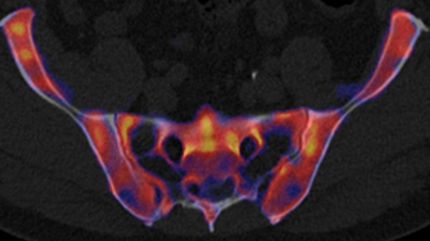 xSPECT-CT pelvis |
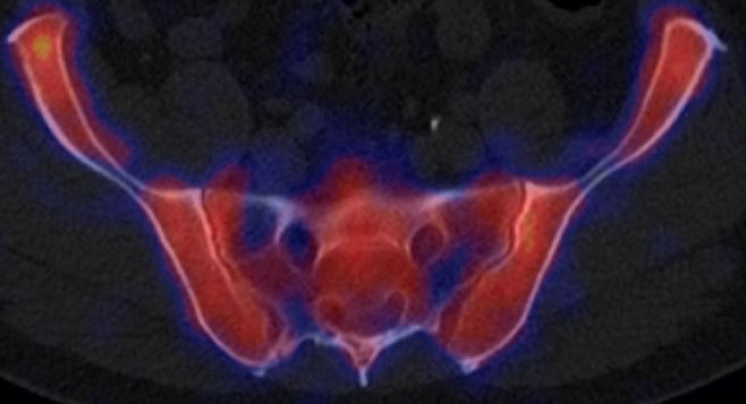 SPECT-CT pelvis |
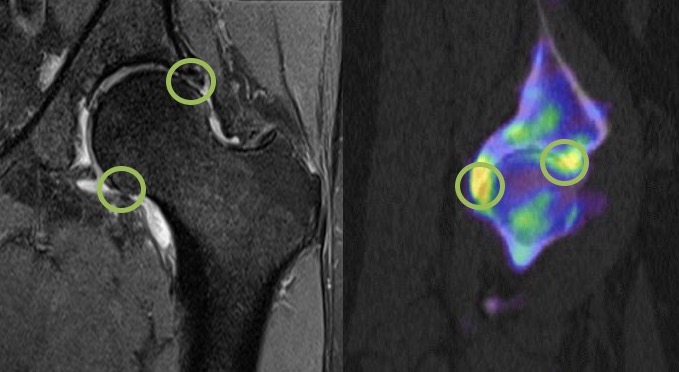 MRI and XSPECT bone |
Discussion
This study agrees with a previous study of reader confidence that indicated xSPECT bone scans offer substantial clinical advantages and reader confidence compared with conventional SPECT bone scans.[i] [ii] The change in both final diagnosis and diagnostic confidence was only a slightly smaller magnitude than my earlier study documenting the change from planar to SPECT-CT bone imaging (unpublished data from 2008 on 524 consecutive cases using the same methodology and presented at the Auckland ANZSNM 2010).
There were very few identifiable problems however, the higher resolution brings a greater amount of data for interpretation. It is therefore more important to correlate the greater number of findings with the patient’s specific clinical setting.
[i] Vija, A. H., Ma, J., Bartenstein, P et al. (2015). ROC analysis for xSPECT Bone. Journal of Nuclear Medicine, 56(supplement 3), 1279–1279.
[ii] Vija, A. H., Ma, J., Bartenstein, P et al. (2014). Reader confidence in lesion detection and lesion characterization with xSPECT Bone. J Nucl Med, 55(supplement 1), Abstract 1499.
*Duncan, I. (Garran Medical Imaging): THE CLINICAL VALUE OF XSPECT VERSUS SPECT IN BONE SCANNING. A HEAD TO HEAD COMPARISON OF 200 CASES. Internal Medicine Journal, April 2017 (Supplement 1), 22–23 (Abstract O60).
**Full publication European Journal of Hybrid Imaging 2018
***For more about xSPECT bone see here.