

Executive Summary
Platelet-Rich Plasma (PRP) represents an increasingly explored autologous orthobiologic in the therapeutic landscape for osteoarthritis (OA). Derived from a patient’s own blood through a centrifugation process, PRP delivers a concentrated solution of platelets and an array of bioactive growth factors, including platelet-derived growth factor (PDGF), transforming growth factor (TGF), insulin-like growth factor (IGF), fibroblast growth factor (FGF), and vascular endothelial growth factor (VEGF).1 These components are believed to stimulate an immunological and inflammatory response that supports physiological healing, modulates inflammation, promotes tissue repair, and stimulates anabolic processes within chondrocytes, synoviocytes, and mesenchymal stem cells, with the overarching aim of alleviating pain and potentially slowing disease progression.1
Current evidence suggests that PRP injections, particularly leukocyte-poor PRP (LP-PRP), can offer superior pain relief and functional improvement when compared to traditional treatments such as hyaluronic acid (HA) and corticosteroids. This benefit is most notable in patients with mild to moderate knee OA (Kellgren–Lawrence grades I–III).2 While some studies indicate sustained effects up to 12 months, it is important to note that the onset of pain relief with PRP may be slower than with corticosteroids.4 The effectiveness of PRP can also vary significantly depending on the specific joint affected.6
The safety profile of PRP is generally favorable, with reported adverse events typically mild and temporary, primarily consisting of post-injection pain.7 The autologous nature of PRP inherently minimizes the risk of immune reactions or disease transmission.10
Despite these promising outcomes, the clinical utility of PRP remains a subject of ongoing discussion. A primary challenge lies in the substantial variability of preparation protocols, including differences in platelet concentration, leukocyte content, and activation status.1 This heterogeneity complicates consistent conclusions and reproducibility across studies. Consequently, major clinical guidelines from prominent organizations such as the American Academy of Orthopaedic Surgeons (AAOS), American College of Rheumatology (ACR), and Osteoarthritis Research Society International (OARSI) maintain cautious or inconclusive stances, citing insufficient or low-quality evidence and concerns regarding study bias.2 Furthermore, current imaging studies do not definitively demonstrate that PRP promotes cartilage regeneration or halts the structural progression of OA.2 The cost-effectiveness of PRP is also a complex issue, with some analyses suggesting it may not be cost-effective from a broad societal perspective, largely due to a lack of established efficacy in definitively delaying total knee arthroplasty (TKA). However, other analyses indicate potential cost-effectiveness from a payer perspective if the total treatment cost remains below certain thresholds over a 12-month period.17

Introduction to Osteoarthritis and Platelet-Rich Plasma
Osteoarthritis (OA) is a pervasive and debilitating chronic degenerative joint disease that impacts millions globally. It is characterized by the progressive breakdown of articular cartilage, inflammation of the synovial membrane, and structural changes in the subchondral bone.6 Patients typically present with localized inflammation, persistent pain, joint stiffness, and restricted joint movement, which collectively diminish their capacity for daily activities and significantly reduce their overall quality of life. The global burden of OA continues to escalate, driven by an aging population and rising rates of obesity.6
Conventional management strategies for OA span a spectrum from conservative approaches, such as physical therapy, non-steroidal anti-inflammatory drugs (NSAIDs), and intra-articular injections (corticosteroids, hyaluronic acid), to surgical interventions reserved for advanced stages. While these treatments aim to alleviate symptoms, they often provide only temporary relief, may be associated with side effects, or are considered for severe, refractory cases.1 The limitations of these traditional methods have spurred interest in biological therapies that leverage the body’s intrinsic healing capabilities.
Platelet-Rich Plasma (PRP) has emerged as a prominent example of such a biological treatment. It is an autologous product, meaning it is derived directly from the patient’s own blood, thereby inherently minimizing the risk of adverse immune reactions or disease transmission associated with allogeneic (donor) products.10 The preparation of PRP involves centrifuging a whole blood sample to achieve a concentration of platelets significantly higher than that found in whole blood.1
The therapeutic rationale underpinning PRP’s use in OA is centered on the rich array of growth factors and cytokines released from activated platelets. These include Platelet-Derived Growth Factor (PDGF), Transforming Growth Factor-beta (TGF-β), Insulin-like Growth Factor (IGF), Fibroblast Growth Factor (FGF), and Vascular Endothelial Growth Factor (VEGF).1 These bioactive factors are believed to foster tissue repair and regeneration through several proposed mechanisms: stimulating angiogenesis (the formation of new blood vessels), promoting cellular migration and proliferation (e.g., of chondrocytes, synoviocytes, and mesenchymal stem cells), and enhancing extracellular matrix deposition.1 Furthermore, PRP is thought to modulate inflammatory and catabolic processes within the joint, thereby shifting the joint environment towards an anabolic (tissue-building) state.20
Despite the strong mechanistic plausibility, where PRP’s growth factors and cytokines are theoretically capable of promoting cell proliferation, matrix synthesis, angiogenesis, and anti-inflammatory effects, this robust biological foundation has not consistently translated into definitive, broadly accepted clinical proof of structural cartilage regeneration or actual disease modification as observed via imaging.1 For instance, while PRP aims to stimulate tissue repair, studies, including the RESTORE trial, have not shown significant differences in cartilage loss between PRP and placebo, indicating that PRP does not definitively halt structural progression of OA.2 This observation highlights a significant gap between the promising
in vitro and theoretical in vivo mechanisms and the actual clinical and structural outcomes. This disconnect suggests that the complex in vivo joint environment may limit PRP’s full regenerative potential, or that current clinical applications or imaging modalities are insufficient to induce or detect meaningful structural changes. This understanding is crucial for guiding future research, emphasizing the need for studies that explore more potent PRP formulations, optimized delivery methods, or combination therapies to truly leverage its regenerative potential. It also underscores the importance of developing more sensitive and objective biomarkers or imaging techniques that can detect subtle cartilage repair, rather than relying solely on macroscopic structural changes. This reframes PRP’s current role as primarily symptomatic, while the “regenerative” aspect remains a significant area for future investigation.
Clinical Efficacy of PRP in Osteoarthritis
The clinical efficacy of PRP in osteoarthritis varies across different joints and depends on the duration of follow-up. While some areas show promising results, others present mixed or inconclusive evidence.
Overall Pain and Functional Outcomes across various affected joints
Knee Osteoarthritis (KOA):
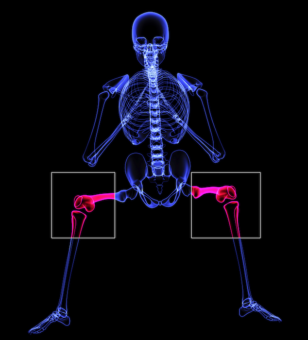
PRP injections, particularly leukocyte-poor PRP (LP-PRP), have demonstrated superior pain relief and functional improvement when compared to hyaluronic acid (HA) and corticosteroids in patients with mild to moderate KOA (Kellgren–Lawrence grades I–III).2 Retrospective analyses of large joint OA, including the knee, have reported clinically significant improvements in pain, with Visual Analog Scale (VAS) scores improving by 49% at 6 months and 45% at 12 months. Functional outcomes, as measured by the Single Assessment Numeric Evaluation (SANE) score, showed improvements of 44% at 6 months and 39% at 12 months.20 Multiple PRP injections have consistently been found to be more effective than a single injection for KOA, suggesting a cumulative benefit.20 The benefits of PRP for KOA typically last between 6 to 12 months, with a reported success rate of 60-70%, defined as at least a 50% improvement in pain and function.5 Meta-analyses of randomized controlled trials (RCTs) generally indicate that PRP performs better than saline injections, particularly when longer-term results are analyzed.5 Overall, PRP has been shown to significantly alleviate pain symptoms (based on VAS and WOMAC scores) and generally improve function, activity levels, participation in sports, quality of life, and joint stiffness within a 12-month follow-up period.19
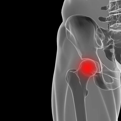
Hip Osteoarthritis (HOA):
Evidence for HOA is less consistent compared to KOA. A systematic review concluded that PRP injection therapy did not significantly reduce pain symptoms in patients with HOA when compared to control groups.6 This lack of significant effect was hypothesized to be due to the hip joint’s deeper cavity, reduced vascularity, and susceptibility to femoral head necrosis, which may hinder the full function of PRP in the hip joint.6 However, a more recent double-blind, randomized controlled trial (conducted from 2019-2021) specifically focusing on hip OA secondary to developmental dysplasia of the hip (DDH) presented contrasting findings. This study reported that PRP was at least as effective as hyaluronic acid (HA), showing a marginally higher improvement in pain-VAS scores (38.5 for PRP vs. 18.7 for HA; P=.041) and significant improvements in WOMAC stiffness scores at 24 weeks compared to HA.22 Another meta-analysis investigating PRP combined with HA for HOA found that dual therapy led to a
higher perception of pain scores at 3 and 12 months compared to PRP alone, suggesting that the addition of HA might paradoxically worsen outcomes for hip OA.23
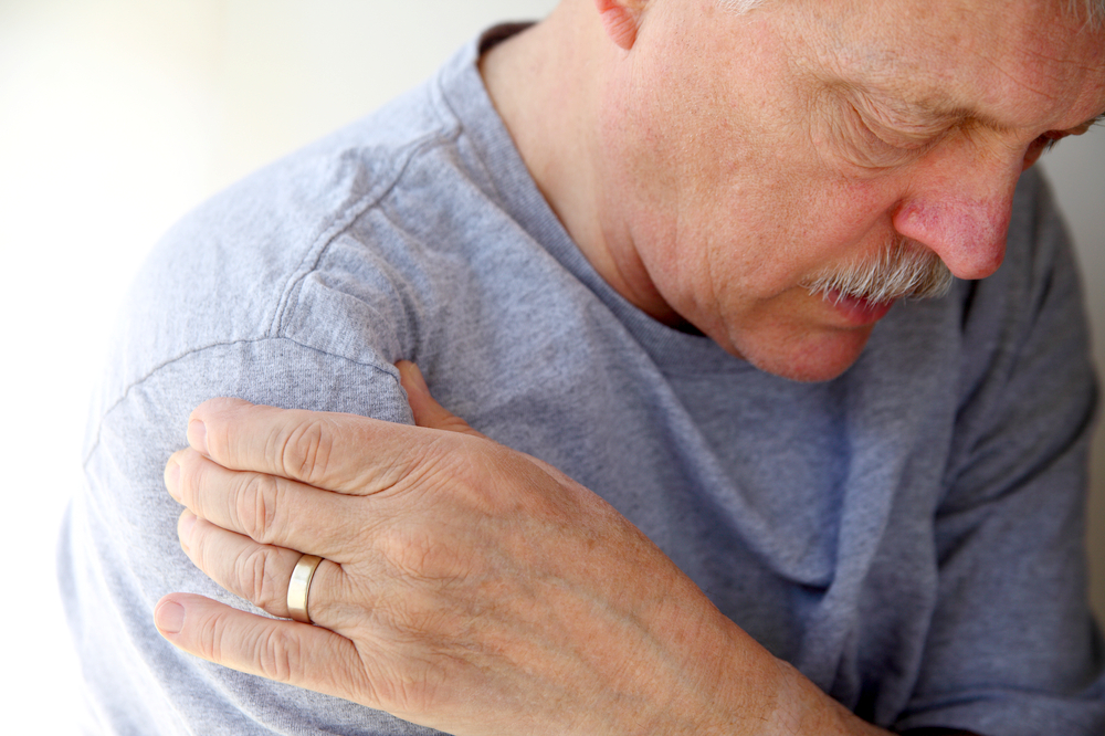
Shoulder Osteoarthritis (AOA) / Rotator Cuff Tendinopathy:
PRP injections have been reported as effective in improving VAS pain scores in ankle osteoarthritis (AOA).6 While not strictly OA, studies on rotator cuff tendinopathy often provide insights into PRP’s effects on joint-related soft tissues. For this condition, PRP significantly reduced pain and improved function in the short term (within 3-6 months) compared to both placebo and corticosteroids.10 A meta-analysis indicated that PRP is a safe and effective intervention for long-term pain control and shoulder function in rotator cuff disorders, with a significant difference in favor of PRP observed at 12 months post-intervention for pain.8
Other Large Joints (Elbow, Ankle, TMJ):
PRP injections were also found to be effective in improving VAS pain scores for temporomandibular joint osteoarthritis (TMJOA).6 For lateral epicondylitis (a common elbow tendinopathy), PRP demonstrated superior early and mid-term clinical outcomes compared to glucocorticoid and saline, showing sustained pain reduction and better functional assessments.25
The efficacy of PRP appears to be highly dependent on the specific joint, suggesting that anatomical factors, such as joint depth, vascularity, and mechanical loading, as well as the specific underlying pathology (e.g., tendinopathy versus true articular cartilage degeneration), significantly influence treatment success. The observation that outcomes for hip OA are less consistent and are attributed to anatomical challenges, such as the hip joint’s deeper cavity and reduced vascularity, underscores that the local microenvironment and the accessibility for effective delivery are critical determinants of PRP’s success. This highlights the necessity for joint-specific research rather than generalizing findings from one joint to another. It also suggests that future research should focus on optimizing PRP formulations and injection techniques tailored to the unique biomechanical and biological characteristics of each affected joint. Clinically, this means practitioners should adjust expectations and treatment recommendations based on the specific joint involved and the available evidence for that site.
Analysis of short-term versus long-term benefits
For knee OA, the benefits of PRP typically manifest and can last for 6 to 12 months.5 For rotator cuff tendinopathy, while significant short-term benefits (pain reduction, functional improvement within 3-6 months) are observed, long-term benefits (beyond 12 months) appear limited, with retear rates and function becoming similar to control groups.10 A systematic review specifically for chronic tendinopathy (encompassing various anatomical sites) found that PRP significantly reduced pain at both 6 and 12 months. Limited data also suggested this effect extended to 24 months, though heterogeneity in outcome measures precluded a pooled analysis of functional improvement.26
Table 1: Summary of PRP Efficacy Outcomes (Pain & Function) by Joint and Follow-up Duration
| Joint | Primary Outcome(s) Assessed | Key Findings (PRP vs. Comparator) | Comparator(s) | Follow-up Duration | Relevant Sources |
| Knee Osteoarthritis (KOA) | Pain (VAS, WOMAC, NPRS), Function (WOMAC, KOOS, SANE) | Superior pain relief & functional improvement vs. HA & CS (mild-mod OA); 49% VAS, 44% SANE improvement at 6 mos; 60-70% success rate (50% pain/function improvement) for 6-12 mos. Multiple injections > single. | HA, CS, Saline, BMAC | Short-term (<6 mos), Medium-term (6-12 mos), Long-term (>12 mos) | 2 |
| Hip Osteoarthritis (HOA) | Pain (VAS), Function (WOMAC) | Inconsistent. Some studies: No significant pain reduction vs. controls. Other studies (DDH-related OA): Marginally higher pain-VAS improvement vs. HA; significant WOMAC stiffness improvement vs. HA at 24 weeks. HA addition to PRP may worsen pain. | HA, Control | Short-term (<6 mos), Medium-term (6-12 mos) | 6 |
| Shoulder/Rotator Cuff Tendinopathy (RCT) | Pain, Function (SPADI, DASH, ASES, CMS) | Significant short-term pain relief & functional improvement vs. placebo/CS (within 3-6 mos). Safe & effective for long-term pain control & shoulder function (12 mos). Limited long-term structural benefits. | Placebo, CS | Short-term (<6 mos), Medium-term (6-12 mos), Long-term (>12 mos) | 10 |
| Lateral Epicondylitis (LE) | Pain (VAS), Function (DASH, PRTEE) | Superior early & mid-term clinical outcomes vs. glucocorticoid & saline; sustained pain reduction & better functional assessments. | Glucocorticoid, Saline | Short-term (<6 mos), Medium-term (6 mos) | 25 |
| Ankle Osteoarthritis (AOA) | Pain (VAS) | Effective in improving VAS pain scores vs. controls. | Control | Not specified | 6 |
| Temporomandibular Joint Osteoarthritis (TMJOA) | Pain (VAS) | Effective in improving VAS pain scores vs. controls. | Control | Not specified | 6 |
Comparative Effectiveness of PRP in Osteoarthritis
The comparative effectiveness of PRP against other non-operative treatments for OA reveals distinct profiles, often highlighting trade-offs between immediate relief and sustained benefits.
PRP versus Hyaluronic Acid (HA) injections
For knee OA, PRP generally appears to outperform HA in most studies, with its benefits often lasting longer, typically 6 to 12 months, compared to HA’s typically shorter duration of effect.2 A meta-analysis of 42 randomized controlled trials (RCTs) specifically comparing HA and PRP for KOA found that PRP resulted in significantly lower Visual Analog Scale (VAS) and Western Ontario and McMaster Universities Arthritis Index (WOMAC) scores, indicating superior performance across various evaluation indices.3 Studies comparing multiple injections have also shown PRP to be superior; for instance, multiple PRP injections resulted in an 80% reduction in VAS scores for KOA after 12 weeks, compared to a 60% reduction with multiple HA injections.21 Furthermore, PRP has demonstrated a significantly higher rate of successful outcomes and lower rates of reintervention when compared with HA in meta-analyses.30
However, for hip OA, the evidence is more mixed. While one study found PRP to be at least as effective as HA, and even marginally superior in pain-VAS scores for OA secondary to developmental dysplasia of the hip 22, another meta-analysis suggested that the addition of HA to PRP for hip OA might paradoxically worsen pain outcomes.23
PRP versus Corticosteroid (CS) injections
The comparison between PRP and CS injections for OA presents a nuanced picture, often involving a trade-off between speed of onset and duration of effect. For knee OA, CS injections can produce better initial pain relief, particularly within the first 4 to 6 weeks post-treatment.5 However, PRP typically outperforms CS at 3 to 6 months after treatment, providing more sustained improvements in pain and function.2 A recent prospective, double-blinded RCT (2025) specifically for mild to moderate KOA found that CS injections provided significantly greater pain reduction at 6 weeks and met the minimal clinically important difference (MCID), whereas PRP did not. At 3 months, improvements were similar between both groups, but neither met MCID. This particular study suggests tempering expectations regarding PRP’s short-term pain relief.4 Conversely, a systematic review and meta-analysis concluded that PRP was significantly better than CS in reducing knee OA symptoms (pain, stiffness, functionality) at 3, 6, and 9 months post-intervention, with benefits observed up to 12 months.27 For rotator cuff tendinopathy, PRP has been shown to significantly reduce pain and improve function in the short term compared to CS. A study found PRP more effective than CS, providing faster and more sustained improvements in pain, functional recovery, and range of motion (ROM), along with fewer adverse events.10 Similarly, for lateral epicondylitis, PRP demonstrated superior early and mid-term clinical outcomes compared to glucocorticoid (CS) and saline, showing sustained pain reduction.25
There appears to be a consistent pattern where corticosteroids offer faster, acute pain relief, while PRP, if effective, tends to provide more sustained, albeit slower-onset, benefits. This difference in temporal efficacy aligns with their distinct mechanisms of action: corticosteroids primarily act as potent anti-inflammatories, providing rapid symptomatic relief, while PRP’s action involves a more complex biological cascade of growth factors and cellular modulation, which takes time to initiate and sustain tissue-level changes. Crucially, repeated corticosteroid injections may have detrimental effects on articular cartilage 20, a risk not associated with PRP. This implies a strategic choice in treatment: for acute pain flare-ups or situations requiring rapid return to activity, CS might be preferred. For chronic management, aiming for more durable effects without the risk of cartilage degradation, PRP could be a more suitable option. This understanding is vital for patient counseling, as it sets realistic expectations regarding the onset and duration of benefits for each treatment. Clinicians can leverage PRP’s favorable long-term safety profile as a key advantage over repeated CS injections. It also opens the door for potential sequential or combination therapies, where an initial CS injection could manage acute pain, followed by PRP to foster more sustained biological effects and potentially delay the need for surgery.
PRP versus Placebo/Saline
Meta-analyses of RCTs for knee OA generally indicate that PRP injections perform better than saline injections, particularly when considering longer-term results.5 However, high-quality placebo-controlled trials have shown inconsistent long-term benefits for KOA, with some failing to demonstrate superiority over saline beyond 6-12 months.2 For chronic tendinopathy generally, a systematic review found that in the majority of comparisons, the effect size of US-guided PRP injection was comparable to control groups (including saline, steroid, etc.), with no clear between-group differences in pain and functional measures for various tendinopathies.7
PRP versus Physical Therapy and other conservative management strategies
PRP is often proposed as an alternative or an improvement upon existing nonpharmacologic therapies, such as exercise and physical therapy, as well as analgesics and anti-inflammatory agents.31 For patellar tendinopathy, physical therapy (especially eccentric training) remains the cornerstone of conservative treatment. While PRP is considered promising, some studies indicate that eccentric exercise therapies yield the best short-term results, whereas multiple PRP injections may provide superior long-term outcomes.32 A retrospective analysis of large joint OA patients who received PRP combined with a rehabilitation program demonstrated clinically significant improvements in pain and function, with active rehabilitation identified as a favorable prognostic factor.20 For rotator cuff tendinopathy, where traditional treatments like physical therapy can provide inconsistent relief, PRP has emerged as a promising regenerative therapy.10
PRP versus Bone Marrow Aspirate Concentrate (BMAC)
Studies directly comparing PRP to Bone Marrow Aspirate Concentrate (BMAC) for knee OA have generally found no significant difference in pain scores or functional outcomes at various follow-up points (e.g., 6, 12, or 24 months).5 There is currently no definitive human study providing strong evidence that BMAC regrows cartilage or offers superior benefits compared to PRP in the treatment of knee OA.5
PRP as an alternative to surgical interventions
PRP is often positioned as a minimally invasive alternative to surgical procedures for chronic tendinopathies and various forms of OA.10 For end-stage OA, joint replacement remains the standard treatment, and intra-articular injections, including PRP, are primarily utilized as a buffer to postpone operative management.29 However, for tendinopathy, a single open-label randomized controlled trial failed to demonstrate a statistically significant reduction in the need for surgical revision with the addition of PRP.31 Furthermore, a cost-effectiveness analysis for knee OA found that PRP was not cost-effective from a societal perspective, largely due to a lack of established efficacy in delaying total knee arthroplasty (TKA).17
Table 2: Comparative Efficacy of PRP vs. Other Non-Operative Treatments for OA
| Comparator | Joint(s) Studied | PRP Performance vs. Comparator | Key Findings/Nuances | Duration of Effect Noted | Relevant Sources |
| Hyaluronic Acid (HA) | Knee OA, Hip OA | Superior for KOA, mixed for HOA | Superior pain/function for KOA; benefits often last 6-12 mos. Multiple PRP injections > multiple HA injections for KOA. For HOA, PRP may be at least as effective as HA, but HA addition to PRP may worsen pain. | Medium-term (6-12 mos) | 2 |
| Corticosteroids (CS) | Knee OA, Rotator Cuff Tendinopathy, Lateral Epicondylitis | Superior long-term for KOA/RCT/LE, inferior short-term for KOA | CS offers faster initial pain relief (4-6 weeks for KOA), but PRP typically outperforms at 3-6 months and provides more sustained benefits. PRP has fewer adverse events for RCT. | Short-term (<6 mos), Medium-term (3-6 mos), Long-term (>12 mos for some RCT) | 10 |
| Placebo/Saline | Knee OA, Chronic Tendinopathy | Generally superior for KOA long-term, comparable for some tendinopathies | PRP often performs better for KOA, especially long-term. Inconsistent long-term benefits for KOA in some high-quality trials. Effect size comparable for many tendinopathies. | Short-term (<6 mos), Medium-term (6-12 mos), Long-term (>12 mos) | 7 |
| Physical Therapy (PT) | Patellar Tendinopathy, Large Joint OA, Rotator Cuff Tendinopathy | Complementary/Superior long-term for some tendinopathies | PT remains mainstay for patellar tendinopathy (eccentric training best short-term). PRP combined with rehabilitation shows significant improvements for large joint OA. PRP is a promising alternative when PT provides inconsistent relief for RCT. | Varies, often long-term | 10 |
| Bone Marrow Aspirate Concentrate (BMAC) | Knee OA | Similar outcomes | No significant difference in pain/function between PRP and BMAC at 6, 12, or 24 months. No strong evidence that BMAC regrows cartilage or is superior to PRP. | Medium-term (6-24 mos) | 5 |
| Surgical Interventions | Chronic Tendinopathy, End-stage OA | Alternative to postpone surgery; not always reduce need for revision | PRP is minimally invasive alternative for chronic tendinopathies/OA. Used to postpone joint replacement for end-stage OA. RCT failed to show reduction in surgical revision for tendinopathy. Not cost-effective for delaying TKA from societal perspective. | Long-term | 10 |
Safety Profile and Adverse Events of PRP in OA
Platelet-Rich Plasma (PRP) is generally considered a safe intervention for osteoarthritis, characterized by a low incidence of serious adverse events.2 The autologous nature of PRP, being derived from the patient’s own blood, inherently minimizes the risk of adverse reactions, immune responses, or disease transmission that could be associated with non-autologous treatments.10
The most frequently reported adverse event among patients receiving PRP injections is temporary post-procedural pain at the injection site.8 This discomfort is typically mild and transient, usually resolving within a few days. In a systematic review of ultrasound (US)-guided PRP for various tendinopathies, no serious adverse events, such as application site infections or severe inflammatory reactions, were reported in either the PRP or control groups over follow-up periods ranging from 1 to 24 months.7 However, less serious, short-term adverse events, predominantly post-injection pain, were observed to be more common among PRP recipients compared to control groups receiving other US-guided injections (e.g., saline, steroid, lidocaine, or bone marrow-mesenchymal stem cells). The Visual Analog Scale (VAS) scores for pain associated with the injection itself also favored the control groups.7
When comparing PRP to hyaluronic acid (HA) for knee OA, the incidence of adverse events was noted to be higher in the PRP group, though all reported complications were mild.3 Conversely, for rotator cuff tendinopathy, PRP therapy was associated with a significantly lower incidence of overall adverse events compared to corticosteroid injections, with fewer cases of mild pain at the injection site and no infections reported in the PRP group.28 Only one study on rotator cuff tendinopathy reported other adverse effects such as frozen shoulder and tear extension, but generally, PRP is considered a safe treatment alternative.8
While PRP is consistently characterized as safe due to its autologous nature, a closer examination reveals that transient post-injection pain is a common and even expected side effect. This minor discomfort, however, needs to be weighed against the potential for more sustained benefits and the absence of the long-term cartilage-damaging effects associated with some other treatments. The transient post-injection pain observed with PRP is likely a physiological manifestation of the initial inflammatory and healing cascade triggered by the concentrated growth factors.1 This implies that a certain degree of discomfort is part of the therapeutic process, rather than a purely negative side effect. This contrasts with corticosteroids, which offer rapid pain relief by suppressing inflammation but may have long-term detrimental effects on articular cartilage 20, a risk not cited for PRP. Therefore, the choice involves a trade-off: immediate, potent anti-inflammatory relief with potential long-term tissue risks (corticosteroids) versus a slower onset of relief accompanied by initial discomfort, but with a more favorable long-term safety profile and potential for tissue modulation (PRP). This nuanced understanding of PRP’s safety profile is critical for effective patient counseling. Patients should be explicitly informed about the likelihood of temporary post-injection pain, framing it as an expected part of the biological response. This also strengthens the argument for PRP as a potentially preferable option for long-term management, especially when considering the cumulative risks of repeated corticosteroid injections, allowing clinicians to position PRP as a safer, regenerative-focused alternative.
Factors Influencing PRP Efficacy and Outcomes in OA
The efficacy and outcomes of PRP in osteoarthritis are significantly influenced by a multitude of factors, primarily related to the preparation methods, the injection regimen, the severity of the osteoarthritis, and patient-specific characteristics. The pervasive lack of standardization in PRP preparation and application is a critical underlying factor that consistently undermines definitive conclusions and broad clinical adoption.
Variability in PRP preparation methods
A significant challenge and a primary reason for inconsistencies in patient outcomes and the practicality of PRP clinical applications stem from the large number and inherent variability of commercially available PRP and PRP-like systems.34 These systems differ substantially in blood collection volumes, centrifugation parameters (speed and time), and overall preparation protocols, which in turn lead to distinctive PRP properties and bioformulations. The absence of a clear consensus on standardizing PRP preparation protocols and adequate reporting on bioformulations in clinical applications further complicates therapeutic analysis and reproducibility.34 This inherent heterogeneity makes it exceedingly difficult to compare results across studies, replicate findings consistently, or establish a universally effective “PRP” product. This lack of standardization directly causes the cautious or negative recommendations issued by major clinical guidelines like AAOS, ACR, and OARSI.2 These authoritative bodies cannot endorse a treatment where the “active ingredient” is ill-defined or irreproducible, as it violates the principles of evidence-based medicine and patient safety/predictability. The “risk of bias” and “low-quality evidence” cited by these guidelines are often direct consequences of this methodological inconsistency. The absence of standardized protocols and the subsequent lack of strong guideline endorsement create significant barriers to widespread clinical utility, limit consistent reimbursement by healthcare payers 2, and perpetuate the ongoing discussion about PRP’s true value.1 This suggests that the future of PRP in OA hinges not just on demonstrating efficacy, but on achieving rigorous standardization to enable robust, reproducible clinical outcomes and clear regulatory pathways.
Platelet Concentration and Dosing: The therapeutic effects of PRP are primarily attributed to the release of growth factors from activated platelets. While it is generally assumed that higher platelet dosages will lead to a greater concentration of released bioactive factors, research indicates that the correlation between platelet dose, concentration, and the release of growth factors may not be consistently controlled due to variations in baseline platelet counts among individuals and differences in preparation methods.34 Furthermore,
in vitro studies suggest that very high concentrations of growth factors are not necessarily advantageous and may even be counterproductive due to receptor saturation.34 Effective clinical PRP is often characterized by a minimum platelet concentration of more than 1 × 10^6/µL or approximately a five-fold increase from baseline, though the optimal concentration for OA remains a subject of ongoing debate.34
Leukocyte Content (Leukocyte-Poor PRP vs. Leukocyte-Rich PRP): The presence and concentration of leukocytes in PRP preparations vary significantly depending on the processing device and protocol.34 Leukocytes are known to influence wound healing and tissue repair, but their pro-inflammatory and immunologic effects can potentially negate the beneficial effects of PRP by causing cell and tissue damage.1 For intra-articular treatment of KOA, leukocyte-poor PRP (LP-PRP) is generally preferred, although the literature remains controversial regarding the optimal leukocyte content.1 A systematic review found that LP-PRP with a high platelet concentration (without an activator) was most effective for improving WOMAC function and stiffness scores at 12-18 months for OA.12 Conversely, LR-PRP with a low platelet concentration (using both anticoagulant and activator) showed the greatest efficacy for WOMAC pain and stiffness scores within 6 months.12 The role of leukocytes is complex; some studies suggest positive effects (e.g., antibacterial action, immune microenvironment regulation), while others point to negative effects (e.g., release of matrix metalloproteinases [MMPs], increased pro-inflammatory cytokines).36 LR-PRP might induce a more severe inflammatory response but potentially stimulate faster tissue repair in early tendinopathy, whereas LP-PRP may increase anti-inflammatory mediators and promote anabolism in later stages.36 For lateral epicondylitis, leukocyte-rich PRP has been suggested to provide more robust and durable pain relief.25
Red Blood Cell (RBC) Content: The presence of red blood cells in PRP specimens can be detrimental. When RBCs are damaged (e.g., during phlebotomy or inadequate centrifugation), they release toxic hemoglobin and iron, leading to oxidative stress, increased cell apoptosis, and potentially cartilage degeneration.34 Therefore, minimizing or eliminating RBC contamination from PRP formulations is considered crucial for optimizing therapeutic outcomes.34
Activation Protocols: PRP must be activated to release the growth factors stored within platelet granules. Common additive agents for activation include calcium chloride and/or thrombin.1 The choice of activator and the duration of activation significantly influence the rate and profile of growth factor release. Bovine thrombin, for instance, causes a faster release of growth factors but is generally not recommended due to toxicity concerns.36 Calcium chloride is considered safer and leads to a slower, more sustained release over several days.36 Photoactivation has also been explored, demonstrating sustainable growth factor release over a longer period.36 While a recent meta-analysis indicated that exogenously activated PRP is more effective in improving pain and function for knee OA, it remains unclear whether rapid or sustained delivery of growth factors is ideal for optimal wound healing.1
Injection regimen
Number of Injections: Studies suggest that multiple PRP injections are generally more effective than a single injection for knee OA.20 For example, triple injections of PRP, typically separated by a week, were found to be superior to single injections over a 12-month follow-up period for knee OA.27
Volume and Site: The total volume of PRP injected can vary (e.g., 2-4 ml for rotator cuff tendinopathy).8 The specific anatomical area of injection (e.g., osteotendinous junction, tendon, myotendinous junction) can also influence outcomes.36 Ultrasound guidance is frequently employed to ensure accurate delivery of the PRP to the lesion site, which is crucial for efficacy.36
Severity of Osteoarthritis (e.g., Kellgren-Lawrence grades)
The therapeutic benefits of PRP appear to be most pronounced in patients with mild to moderate knee OA, typically corresponding to Kellgren-Lawrence grades I-III or 2-3.2 Emerging evidence suggests that optimal PRP protocols should be tailored to the disease stage. Specifically, low platelet concentration, high leukocyte PRP (e.g., RPRP_LPC_Y_Y) is recommended for early OA due to its anti-inflammatory effects and immediate clinical efficacy. Conversely, high platelet concentration, low leukocyte PRP (e.g., PPRP_HPC) is preferred for more advanced OA to promote tissue repair and regeneration.12
Patient-specific factors
Activity Level: A patient’s activity level, particularly high sports activity (especially at a competition level), has been identified as a favorable prognostic factor for outcomes in large joint OA, including the knee, hip, shoulder, and ankle.20 This may be attributed to higher baseline physical fitness, better muscle strength, and improved proprioceptive abilities in competitive athletes, which could optimize the biological environment for PRP’s effects.20
Concomitant Rehabilitation: Active rehabilitation programs, when combined with PRP injections, have been shown to lead to significant improvements in pain and function for patients with large joint OA. Active rehabilitation is considered a favorable prognostic factor.20
Age: While not definitively conclusive, some findings suggest that younger, more active patients may derive greater benefit from PRP therapy, as it aims to promote tissue regeneration and healing. In contrast, older individuals or those with more advanced degenerative changes might find corticosteroid injections more beneficial in managing symptoms.28
Conclusions
The review of Platelet-Rich Plasma (PRP) in osteoarthritis management reveals a complex and evolving therapeutic landscape. While PRP holds significant promise as an autologous orthobiologic, its widespread adoption and definitive clinical recommendations are currently hampered by several critical factors.
PRP demonstrates a generally favorable safety profile, with transient post-injection pain being the most common adverse event. This temporary discomfort is likely a manifestation of the initial biological cascade triggered by the injection, differentiating it from the potential long-term cartilage-damaging effects associated with repeated corticosteroid injections. This safety profile positions PRP as a potentially attractive option for chronic OA management, particularly when considering the cumulative risks of other interventions.
In terms of efficacy, PRP shows notable benefits for pain relief and functional improvement, particularly in mild to moderate knee osteoarthritis, often outperforming hyaluronic acid and providing more sustained benefits than corticosteroids. However, the onset of pain relief with PRP can be slower than with corticosteroids, suggesting that these therapies may occupy different therapeutic niches – corticosteroids for acute symptom management and PRP for more durable, tissue-modulating effects. The effectiveness of PRP is also highly joint-dependent, with less consistent outcomes observed for hip osteoarthritis compared to the knee, likely due to anatomical constraints and varying pathological mechanisms. This highlights the need for joint-specific research and tailored treatment approaches.
A significant challenge that consistently undermines definitive conclusions and broad clinical adoption is the pervasive lack of standardization in PRP preparation methods. Variations in platelet concentration, leukocyte content, and activation protocols across different commercial systems and clinical studies lead to heterogeneous products and inconsistent patient outcomes. This methodological variability directly contributes to the cautious or inconclusive recommendations from major clinical guidelines, as robust, reproducible evidence is difficult to establish. The absence of strong guideline endorsement, in turn, impacts clinical utility and consistent reimbursement.
Furthermore, despite strong mechanistic plausibility for tissue repair and regeneration, current high-quality imaging studies do not definitively show that PRP promotes cartilage regeneration or halts the structural progression of OA. This discrepancy between theoretical potential and observable structural changes indicates a gap in current understanding or application, suggesting a need for further research into optimized formulations, delivery methods, or more sensitive biomarkers to truly harness PRP’s regenerative capabilities.
In conclusion, PRP is a safe and promising treatment for osteoarthritis, particularly for knee OA, offering sustained symptomatic relief and functional improvement. However, its full potential remains to be unlocked through rigorous standardization of preparation protocols, targeted research addressing joint-specific nuances, and studies that can conclusively demonstrate structural disease modification. Until these challenges are addressed, patient selection, realistic expectation setting, and careful consideration of PRP’s comparative advantages and limitations against other available therapies are paramount in clinical practice.
Works cited
- Platelet-Rich Plasma for Osteoarthritis in 2024 – More Hype, accessed on June 30, 2025, https://www.sportsmed.org/membership/sports-medicine-update/spring-2024/platelet-rich-plasma-for-osteoarthritis-in-2024
- Platelet-Rich Plasma for Knee Osteoarthritis: A Comprehensive …, accessed on June 30, 2025, https://pmc.ncbi.nlm.nih.gov/articles/PMC12156035/
- Comparison of hyaluronic acid and platelet-rich plasma in knee …, accessed on June 30, 2025, https://pubmed.ncbi.nlm.nih.gov/40069655/
- Platelet-Rich Plasma Injections Are Inferior to Corticosteroid …, accessed on June 30, 2025, https://pubmed.ncbi.nlm.nih.gov/40081606/
- Analyzing the performance of platelet-rich plasma and bone marrow aspirate concentrate injections for the treatment of knee osteoarthritis – Mayo Clinic, accessed on June 30, 2025, https://www.mayoclinic.org/medical-professionals/physical-medicine-rehabilitation/news/analyzing-the-performance-of-platelet-rich-plasma-and-bone-marrow-aspirate-concentrate-injections-for-the-treatment-of-knee-osteoarthritis/mqc-20578651
- (PDF) Efficacy and safety of platelet-rich plasma injections for the treatment of osteoarthritis: a systematic review and meta-analysis of randomized controlled trials – ResearchGate, accessed on June 30, 2025, https://www.researchgate.net/publication/371930900_Efficacy_and_safety_of_platelet-rich_plasma_injections_for_the_treatment_of_osteoarthritis_a_systematic_review_and_meta-analysis_of_randomized_controlled_trials
- Ultrasound-guided injection of platelet-rich plasma for …, accessed on June 30, 2025, https://pmc.ncbi.nlm.nih.gov/articles/PMC10072988/
- Platelet-rich plasma for rotator cuff tendinopathy: A systematic …, accessed on June 30, 2025, https://journals.plos.org/plosone/article?id=10.1371/journal.pone.0251111
- Comparison of hyaluronic acid and platelet-rich plasma in knee osteoarthritis: a systematic review – PubMed, accessed on June 30, 2025, https://pubmed.ncbi.nlm.nih.gov/40069655
- Effectiveness of Platelet-Rich Plasma in Treating Rotator Cuff Tendinopathy: A Systematic Review and Meta-Analysis | Journal of Orthopaedic Case Reports, accessed on June 30, 2025, https://jocr.co.in/wp/2025/03/01/effectiveness-of-platelet-rich-plasma-in-treating-rotator-cuff-tendinopathy-a-systematic-review-and-meta-analysis/
- Exploring Platelet-Rich Plasma Therapy for Knee Osteoarthritis: An In-Depth Analysis, accessed on June 30, 2025, https://www.mdpi.com/2079-4983/15/8/221
- The efficacy of platelet-rich plasma preparation protocols in the …, accessed on June 30, 2025, https://pmc.ncbi.nlm.nih.gov/articles/PMC12186406/
- PRP Injection Preparation and Composition – Arthritis-health, accessed on June 30, 2025, https://www.arthritis-health.com/treatment/injections/prp-injection-preparation-and-composition?s=weight
- Management of Osteoarthritis of the Knee (Non-Arthroplasty) – American Academy of Orthopaedic Surgeons, accessed on June 30, 2025, https://www.aaos.org/globalassets/quality-and-practice-resources/osteoarthritis-of-the-knee/oak3cpg.pdf
- (PDF) A Systematic Review of Current Clinical Practice Guidelines …, accessed on June 30, 2025, https://www.researchgate.net/publication/354267185_A_Systematic_Review_of_Current_Clinical_Practice_Guidelines_on_Intra-articular_Hyaluronic_Acid_Corticosteroid_and_Platelet-Rich_Plasma_Injection_for_Knee_Osteoarthritis_An_International_Perspective
- Osteoarthritis Treatment Guidelines from Six Professional Societies: Similarities and Differences – PMC, accessed on June 30, 2025, https://pmc.ncbi.nlm.nih.gov/articles/PMC9377706/
- The Cost-Effectiveness of Platelet-Rich Plasma Injections for Knee …, accessed on June 30, 2025, https://www.researchgate.net/publication/341571517_The_Cost-Effectiveness_of_Platelet-Rich_Plasma_Injections_for_Knee_Osteoarthritis_A_Markov_Decision_Analysis
- What Is the Appropriate Price for Platelet-Rich Plasma Injections for …, accessed on June 30, 2025, https://pubmed.ncbi.nlm.nih.gov/32061971/
- Comprehensive Summary of the Meta-Analyses and Systematic Reviews on Platelet-Rich Plasma Therapies for Knee Osteoarthritis | Military Medicine | Oxford Academic, accessed on June 30, 2025, https://academic.oup.com/milmed/article/189/11-12/e2347/7616422
- Platelet-rich plasma treatment for large joint osteoarthritis: retrospective study highlighting a possible treatment protocol with long-lasting stimulation of the joint with an adequate dose of platelets – PMC, accessed on June 30, 2025, https://pmc.ncbi.nlm.nih.gov/articles/PMC12023473/
- Clinical Outcome of Multiple Platelet-Rich Plasma Injection and Correlation with PDGF-BB in the Treatment of Knee Osteoarthritis – PubMed Central, accessed on June 30, 2025, https://pmc.ncbi.nlm.nih.gov/articles/PMC10890090/
- Effectiveness of platelet-rich plasma in pain management of …, accessed on June 30, 2025, https://academic.oup.com/jhps/advance-article/doi/10.1093/jhps/hnaf008/8058728
- Platelet-rich plasma with versus without hyaluronic acid for hip osteoarthritis: a systematic review and meta-analysis – PubMed, accessed on June 30, 2025, https://pubmed.ncbi.nlm.nih.gov/40206824/
- Effectiveness of Platelet-Rich Plasma in Treating Rotator Cuff Tendinopathy: A Systematic Review and Meta-Analysis – PMC, accessed on June 30, 2025, https://pmc.ncbi.nlm.nih.gov/articles/PMC11907138/
- Platelet-Rich Plasma Provides Superior Clinical Outcomes Without …, accessed on June 30, 2025, https://pmc.ncbi.nlm.nih.gov/articles/PMC12113477/
- Platelet-rich plasma (PRP) injections as a second-line treatment in …, accessed on June 30, 2025, https://pubmed.ncbi.nlm.nih.gov/40073277/
- (PDF) Intra-articular platelet-rich plasma injections versus intra …, accessed on June 30, 2025, https://www.researchgate.net/publication/352466323_Intra-articular_platelet-rich_plasma_injections_versus_intra-articular_corticosteroid_injections_for_symptomatic_management_of_knee_osteoarthritis_systematic_review_and_meta-analysis
- Original Research Article – COMPARING THE EFFICACY OF …, accessed on June 30, 2025, https://www.ijmedph.org/Uploads/Volume15Issue1/282.%20[1776.%20IJMEDPH_Rajesh]%201504-1509.pdf
- Are patients with knee osteoarthritis aware that platelet-rich plasma is a treatment option?, accessed on June 30, 2025, https://pmc.ncbi.nlm.nih.gov/articles/PMC10798754/
- Platelet-Rich Plasma Versus Alternative Injections for Osteoarthritis …, accessed on June 30, 2025, https://pubmed.ncbi.nlm.nih.gov/38420745/
- Orthopedic Applications of Platelet-Rich Plasma, accessed on June 30, 2025, https://www.southcarolinablues.com/web/public/brands/medicalpolicy/external-policies/orthopedic-applications-of-platelet-rich-plasma/
- Platelet-Rich Plasma for Patellar Tendinopathy: A Comprehensive Review of Current Literature – ClinMed International Library, accessed on June 30, 2025, http://clinmedjournals.org/articles/jmdt/journal-of-musculoskeletal-disorders-and-treatment-jmdt-9-123.php?jid=jmdt
- Platelet Rich Plasma Injections for Non-Wound Injections (L39058) – CMS, accessed on June 30, 2025, https://www.cms.gov/medicare-coverage-database/view/lcd.aspx?lcdid=39058
- Platelet-Rich Plasma: New Performance Understandings and …, accessed on June 30, 2025, https://www.mdpi.com/1422-0067/21/20/7794
- Platelet-rich plasma with versus without hyaluronic acid for hip osteoarthritis: a systematic review and meta-analysis – Frontiers, accessed on June 30, 2025, https://www.frontiersin.org/journals/bioengineering-and-biotechnology/articles/10.3389/fbioe.2025.1545431/epub
Platelet-rich plasma in the pathologic processes of … – Frontiers, accessed on June 30, 2025, https://www.frontiersin.org/journals/bioengineering-and-biotechnology/articles/10.3389/fbioe.2023.1187974/full