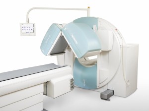
Nuclear medicine encompasses the use of medical radioisotopes to image various parts or systems in the human body. It involves injecting tiny amounts of “radiotracers” into the body to assess the functions of various organs or body systems. It is most commonly used to assess the boney skeleton, the heart, thyroid and kidneys. Unlike x-rays or CT scanner which send x-rays through your body, nuclear medicine collects rays coming from your body. The device used for collection is a gamma camera which turns these emissions into images. More modern technology may combine the use of a gamma camera with a CT (xray) scanner to obtain two sets of information simultaneously. A combined CT (xray) and gamma camera is shown below. The doughnut scanner is the CT and the two square heads are the gamma camera. The bed passes through both scanners in sequence.
The radiotracer will take time to reach or pass through the area of the body to be examined. For example bone scans require 2-4 hours for the radiotracer to be taken up by all the bones. For every tracer and system the timing is different. Most of the radiotracers are based on the isotope technetium which has a short (6 hours) half life and is excreted rapidly by the body after your examination. These timing differences determine the specific scheduling of each examination. Radiotracers do involve tiny doses of radiation and do not cause any side effects during their passage through the body. Most of the compounds used are quickly eliminated from the body.
If you are having a nuclear medicine examination the nuclear medicine technologist will operate the gamma camera and help you through the process.
Many examinations are undertaken in two (or sometimes more) parts. The initial part involves an injection into a vein and sometimes some preliminary pictures. Later more detailed pictures are taken and sometimes a series of images are captures as the tracer passes through the organ system.In some of the test procedures additional activities are undertaken to test the performance of a given organ or system. For example you may be asked to exercise for cardiac testing. You may be given a drug to make your kidneys excrete urine or to maximise blood flow to your heart or brain. Sometimes a test is done before and after such an activity.
The information and images obtained from the examination are compiled and analysed by the nuclear medicine physician and technologist using sophisticated software. The physician will interpret the scans in relation to the particular clinical problem. He or she will often talk to and examine the patient to obtain as much background information as possible.
Finally a report is used regarding the entire procedure. Usually this is a critical input into both the patient’s diagnosis and/or clinical management.
For further information see the AANMS website.

