lateral epicondylitis

Lateral Epicondylitis: A Therapy Update

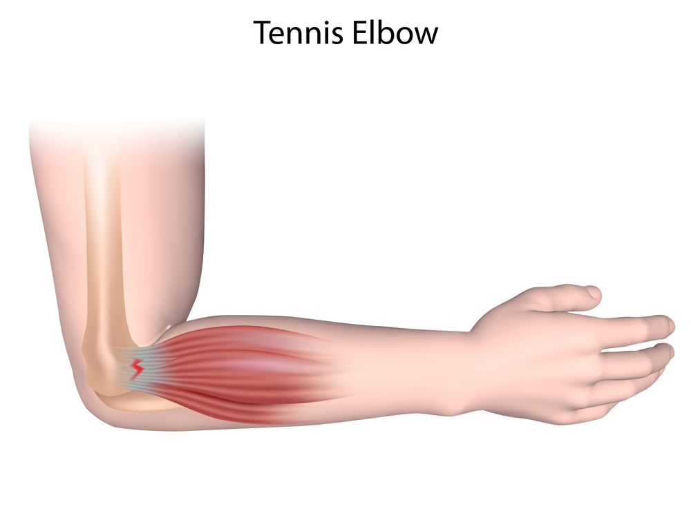
Background  Definition: Lateral epicondylitis is a common, painful condition characterized by pain at the lateral side of the elbow, that increases during gripping or squeezing. Though sometimes referred to as “tennis elbow” it occurs mostly in middle aged adults regardless of sporting activity.  Light microscopy reveals both an excess of fibroblasts and blood vessels that […]

Back pain and Bone scans (updated 2017)

Bone scans of the spine have become a clinically useful part of the evaluation of back pain. Uptake seen on a SPECT-CT correlates with metabolic activity in bone and the increased resolution and specificity of SPECT-CT has allowed a greater number and variety of clinically relevant findings. Most patients with back pain will have an […]

Ultrasound Elastography

Basic principles Recent advances in ultrasound technology have led over several years to the development of elastography. This is the ultrasound equivalent of clinical palpation. There are different types of shear elastic properties which can be assessed by ultrasound. Several types have developed using different deformation forces and different ways of measuring the force. A […]

Nuclear Imaging in Prosthetic Joints

NUCLEAR TRACERS used in imaging prosthetic joints

The tracers discussed in this article are:

Tc-MDP: bone scintigraphy

Tc-sulesomab:  in vivo labelled Fab fragment of IgG1.  While this also binds to neutrophils (5%) and therefore should mimic WBC labelled scans it has a non-specific accumulation at infected sites. 35% of activity at 24hrs is in the bone marrow.

WBC labelled scans: in vitro labelled patient WBC with either Tc or In-111

Gallium 67

FDG (Fludeoxyglucose 18F): is also used for infection imaging but PET scanning is not discussed in this article.


SUMMARY OF OUR APPROACH TO SCANNING PROSTHESIS

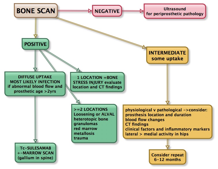
PROSTHETIC JOINT WORKFLOW

WORKFLOW for prosthetic joint complications


[…]

BIRADS BREAST CLASSIFICATION

0 incomplete additional imaging evaluation needed 1 negative   2 benign likelihood of malignancy ~0%. Routine screening recommended 3 probably benign 0-2% likelihood of malignancy. Follow-up in ~6 months 4 suspicious likelihood of malignancy 2-95% 5 highly suspicious >95% risk of malignancy. Requires tissue diagnosis 6 proven malignancy biopsy positive. Surgical excision recommended

Presentations

Imaging of the foot: A presentation to the Australian Sonographers Association  National Conference Canberra May 2012. This briefly covers the forefoot conditions best evaluated/diagnosed with either nuclear medicine or ultrasound. Contrast Enhanced Ultrasound of Liver Lesions This is a 2 slide summary of the behavior of liver lesions to microbubble contrast. Imaging Musculoskeletal Pain A multi-modality talk on […]