
MAY 2013 to DECEMBER 2015 NEWS ARCHIVE
November & December 2015
Most of us are time poor and thinking on this recently I came up with a concept of 10 minute updates for our referrers. You don’t need an exhaustive lecture to get your head around something. We have now progressed the concept to reality and hopefully these short presentations can update referring practitioners on medical imaging from the basics to the most recent technologies, scientific advances, and concepts. It is also an opportunity for forging a better relationship between users and providers of medical imaging, something that is definitely needed. In a little more than half an hour 3 subjects can be covered and there is still plenty of time for informal discussion and chit chat. Kevin Osborn and I launched this on the 2nd December at Garran Medical Imaging. The audience was small (6 including Kevin and I) but you have to start somewhere. After a break in January 2016 we plan to recommence in February, once a month. We could even come to you either in the real or virtual -all requests considered! Just let us know what you would like to hear about (info@driainduncan.com.au). My favorites are, of course, recent advances such as elastography, xSPECT nuclear imaging, PRP therapy and as always any musculoskeletal ultrasound topic.
If you managed to read this sentence I congratulate you and wish you the very best wishes of the season.
October 2015
I have started offering patient’s ultrasound guided platelet rich plasma (PRP) injections at Garran Medical Imaging using a new system from regenlab which fulfils the criteria I have outlined in my update this month. I am confident that this can have a clinical impact where alternatives are extremely limited (steroids, polidocanol, hyaluronate, glucose, autologous blood).
And what about that win against Wales at the World Cup.

Wallabies psyched up for a momentous battle v Wales
September 2015
Some new website additions: New abstract on patellofemoral pain. I was able to attend both the ASUM ultrasound meeting in Sydney and the following week the ASA Special Interest Group meeting, where I gave two workshops, helped in another, and participated in a plenary forum. For those who requested a closer look at some of the images for ankle and foot you can find them here.
For those interested in pulmonary embolism I discovered this useful reference for the diagnosis: the PERC rule to exclude PE if no criteria are present and pre-test probability is <15%.
August 2015
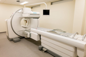
August has been a lot of hard work but great fun. Garran Medical Imaging had a late start in mid July but is now up and running. Plenty of minor troubles but the staff have been amazing and the future looks very bright. There are lots of first for the business which has both embraced principles of customer service from other industries and endeavoured to put the patient at the centre. Needless to say Garran Medical Imaging will be looking at new ways to use cutting edge technology to deliver clinical benefits for patients and doctors. This is my “raison d’être.” Two such technologies are xSPECT bone imaging and shearwave elastography which are feature stories on the website this month. These technologies are currently available at only a few locations in Australia. While xSPECT will raise the bar of nuclear bone imaging, shearwave elastography provides a new way of assessing the liver for those with chronic hepatitis and liver disease.
July 2015
I have teamed up with Dr Kevin Osborn and an enthusiastic team at a new imaging centre of excellence. Garran Medical Imaging is located at Garran Shops (near The Canberra and National Capital Private Hospitals). Garran Medical Imaging aims to be part of the solution for doctors and patients. Kevin and I would like to better partner with referring doctors to help solve clinical problems and improve/de-stress a patient’s journey through the medical system.The practice has been designed to provide excellence at every interaction and from all angles. The patient journey from referral through booking, check-in, scanning, completion and delivery of results has been carefully mapped to ensure a comfortable and stress free experience. We are aiming to make the diagnostic quality of our scans world class.
June 2015
I have commenced a new “ultrasound blog” for the benefit of all the sonographers I work with and those that I don’t. Feel free to send me feedback via email. You can find it under “Sonography grabs” in the categories search menu or via the “Information for Sonographers and Sonologists” section in Resources and Updates.
Garran Medical Imaging is now in the final stages of gestation with equipment partly installed. I am so excited and at the same time terrified of some oversight or further delay (we are only going to open 4 months late). The website will launch this week and our PR campaign is under way. I cannot believe that a medical imaging practice could have had more love and attention prior to debut. Lets hope the momentum rolls on…
May 2015
In conjunction with Dr Kevin Osborn I am proud to announce the creation of Garran Medical Imaging which is a new Canberra Medical Imaging practice designed to deliver best possible service and diagnosis using advanced imaging technology. Together with Nick Ingold and an amazing team we aim to deliver cutting edge diagnostic imaging in a more caring and supportive way. Let us show you why we love our jobs and how we can help make your diagnostic journey the best one possible. Planning has been going on for more than a year and final building construction will be complete in June. The best possible equipment along with truly dedicated staff will open the doors of Garran Medical Imaging for business in July 2015. The website of Garran Medical Imaging will launch soon.
April 2015
I attended the ANZSNM (The Australian and New Zealand Society of Nuclear Medicine) annual Scientific meeting in Brisbane between the 17th and 20th April. As well as getting my dose of educational updates I listened to those who have had the first experience with X-SPECT technology in Australia. This is particularly exciting as Garran Medical Imaging (GMI) will be joining the early adopters of this awesome technology. This aligns with the mantra of GMI which is all about exploring new ways of using medical imaging technology to improve clinical outcome for both doctors and patients. We want to be part of the solution. GMI will be introducing several new technologies into clinical practice and while the technology has been scientifically validated no one is certain how it is best applied or how much clinical difference it will make. I will be encouraging a dedicated team of professionals to collect information that helps deliver solutions. In the meantime check out these images below.
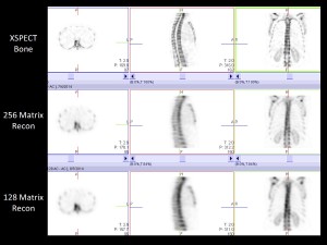
XSPECT BONE v XSPECT v SPECT
The XSPECT bone images on the top row compare with the best current systems on the bottom row. The middle row is the SPECT images on the new system without the X-SPECT processing. These images are prior to fusing with the CT part of the scan. I am sure you can see the difference (click on the image to enlarge).
On another note entirely a new study has shown stretching is unproven in preventing tendon injuries but that footwear is important.
March 2015
Exciting news. A brand new medical imaging practice will be opening in Canberra later this year. This practice has been planned from the ground up in every respect: custom-built modern premises, state of the art equipment and an inclusive team environment. Garran Medical Imaging will have a small and dedicated team of professionals who will set a new benchmark for the industry. Needless to say Iain will be keen to share news and detail over the next few months as the jigsaw comes together. For those professionals who would like to participate a select number of jobs have been advertised here.
February 2015
The website has had a makeover in January -the organisation is different to cope with more content and there are lots of new illustrations. New additions include an abstract about breast tomosynthesis which will hopefully improve the journey for women requiring investigation and screening for breast cancer. This is not yet widely available but will come on stream as radiology centres update their equipment over the next few years.
January 2015
Quiet start to the new year and now accelerating with lots of changes to the website, both in content and organisation. The New Year is also a chance for lots of reading both for technical and spiritual growth. James Alutcher captures some great LIFE TIPS to help us reinvent ourselves in the new year.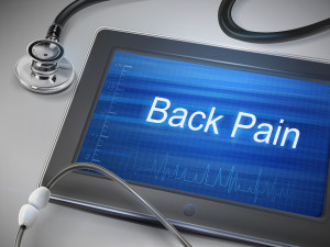
Back pain is such a common problem and to clarify the role of bone scanning and for a quick update check out my latest post.
I have also written a paper on scanning of prosthetic joints (hips, knees, and some back surgeries) with a quick reference flowchart. Unfortunately there are no gold standards for sorting out complications of joint replacement and post-op pain. Nuclear medicine is the next stop after a plain x-ray. See here.
I get a lot of questions about terminology and classification. We all end up using terminology well known to our individual craft groups but not always well understood by others. To tackle this I will progressively add to the website glossaries (found in either the referring doctors area or the Search by category MENU) my commonly used reporting classifications and terms I use, for example stress fractures.
December 2014
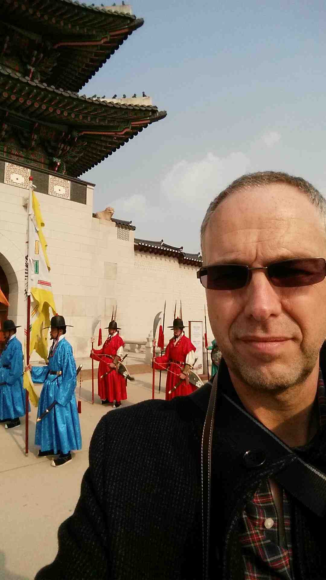
Iain at the Royal Palace Seoul
It was great to get a break and to press the soft reset button. Lots of cool thing will happen in 2015.
November 2014
It was my honor to be invited to join the Samsung Radiology KOL forum. Without any hesitation I have accepted and am off to Seoul to participate with a dozen or so other radiologists from around the world. I am excited about being involved with Samsung in ensuring their new ultrasound is as good as it can be. My impressions so far are that the RS-80 will exceed expectations and I can’t wait to test all its capabilities. I have ordered two for a new and exciting upcoming venture next year. More about that later…
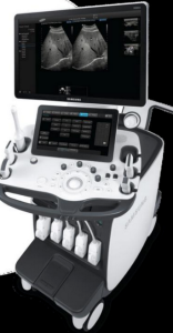
Samsung Medison’s new ultrasound system
Seoul was as far away from Vienna (see October 2013 below) in head space as it is across the globe. Korea is a fascinating place and demonstrates an eagerness to embrace the future that is a little intoxicating. The developers and researches I met demonstrated an optimistic “can do” enthusiasm that suggests Samsung Medical will bring us many innovative solutions (and I was able to witness several in the pipeline). I was able to visit Samsung Medical Centre (a paperless hospital), Samsung Medical Headquarters, its medical imaging research area, and Samsung Electronics headquarters at “Samsung City” in Suwon (35,000 work here). A great and impressive experience. I look forward to working more with Samsung Medical.
September 2014
The web site now contains an embryonic image gallery with a series of foot images being first for publication. This will mostly be useful for those studying in the areas of medical imaging and particularly ultrasound and nuclear medicine. Though SPECT-CT nuclear medicine has become mainstream in Australia its full potential is rarely utilised as there are so many imaging alternatives for any given clinical scenario. Whichever modality is used it is the quality of clinical information that determines how each scan is done and interpreted -so much is missed by looking in the wrong place or ignoring subtle findings which might be critical for that particular case. In ultrasound, apart from looking at the right structures, it is the use of sensitive blood flow techniques, adjuvant tools such as elastography, and clincal skills that are key to getting the most from an examination. In bone scans looking in detail at the right locations, using the right tracer uptake maps and thresholds, the optimum CT windows, and being keenly aware of the precise clinical problem are similarly important. Check out the images.
A recent study shows lung perfusion scintigraphy exposes the pregnant patient and foetus to a lower radiation dose than than CTPA using modern 256-slice CT scanners.
August 2014
Thanks for those who came along to hear me talk at the Canberra ASUM musculoskeletal Ultrasound Workshop. It was the first time I use the concept of the Musculoskeletal Ultrasound PYRAMID. The Pyramid will be developed more and I hope will be a good template for learning and teaching MSK ultrasound. My apologies that the finer points of the concept are still in evolution. Such constructs help us organise our learning and hopefully provide a window into the path forward. Another useful construct or paradigm is the TRIANGLE and the concept of triangulation best explained in this video by Roger James Hamilton.
July 2014
I have just added about a dozen new patient information booklets/downloads to the site (click on the RESOURCES & UPDATES menu) which I hope will be very helpful. I see dozens of such cases every week and I know there is a lot of information to absorb about about a new diagnosis. If you don’t get exactly what you want fell free to send me an email.
I am excited about the latest developments in nuclear medicine technology. Siemens had developed the next generation SPECT-CT system which promises a significant increase in resolution of the combined modality imaging. In addition the system will be quantitative -able to measure tracer uptake per volume. Even now I am thinking up potential clinical applications though it is unlikely I will be able to use such a system for some time. Currently no one has a full system installed in Australia. Could I be the first? Pipe dream at this stage. For those technically minded click here.
June 2014
A new publication involving a comprehensive meta-analysis of studies involving injury prevention was published in the British Journal of Sports Medicine. The authors found that in general terms physical activity was shown to effectively reduce sports injuries. However stretching alone had no beneficial effect. The authors concluded “multiple exposure programmes, proprioception training, and strength training, in that order, showed a tendency towards increasing effect” and “strength training reduced sports injuries to less than one-third”. Unfortunately they noted trials did not separate injuries into acute and repetitive categories which would prove helpful in future studies. From the perspective of evaluating the injuries this may have limited implications but it supports increasing evidence (and my personal bias) that strength training is a critical part of injury rehabilitation.
April 2014
Hamstring injuries are frequent and can be disabling. This new publication from Sweden is an important step in improving our understanding of rehabilitating muscle (specifically hamstring) tears. For me the “Take Home” messages from the study were (in relation to acute hamstring injury): A rehabilitation protocol consisting of mainly lengthening type of exercises (strength and flexibility exercises that primarily involve high loads at long muscle-tendon lengths) is more effective than a conventional protocol. Also from an ULTRASOUND PERSPECTIVE the current study showed that increased recovery time can be expected with (1) pain closer to the ischial tuberosity, or (2) involvement of the free muscle tendon, or (3) oedema close to the ischial tuberosity.
March 2014
Iain presented “Imaging Musculoskeletal Pain” at Regatta Point, Canberra sponsored by National Capital Diagnostic Imaging. This event titled “Imaging guided Injections and Musculoskeletal Imaging” featured Dr Iain Duncan and Dr Nicholas Tsai (Orthopaedic Surgeon). For those who attended this event Iain’s presentation (no audio) can be viewed here. If you would like to give us feedback or suggestions for future events send an email to info@driainduncan.com.au.
February 2014
I often get asked whether a thyroid biopsy is needed in particular patients with multinodular goitre and so I have outlined my approach in this article. The The U.S. Preventive Services Task Force (USPSTF) has published some new recommendations regarding abdominal aortic aneurysm screening.
January 2014
A recent article in Paediatrics concluded that “For children ≤2 years of age with a febrile UTI, an acute DMSA scan is valuable in the exclusion of dilating VUR [vesic-ureteric reflux]. The likelihood of the presence of dilating VUR on MCU is rather low when the result of DMSA is negative. DMSA should be conducted to assess the need for an MCU.” They found the negative predictive value of DMSA was 97.26% for children ≤2 years of age and 100.0% for the older children.
This suggests that a DMSA scan may be a useful tool in infants with a UTI prior to undertaking the more invasive MCU investigations. A positive DMSA is not specific for VUR but a negative one almost always excludes it.
November 2013
I have posted an interesting publication from Morocco on ultrasound findings in patients with ankylosing spondylitis. From my perspective it supports the concept that when I see unexplained enthesitis in the young non-biomechanical causes (such as A.S. and other spondyloarthropathies) need to be considered as diagnostic possibilites.
October 2013
I have just returned from Vienna and the International Atomic Energy Agency where I spent a week interacting and learning from some of the world’s great cardiology imaging gurus at the International Conference on Integrated Medical Imaging in Cardiovascular Diseases. I was both reassured that both in Bega and at NCDI in Deakin our practices are using polished techniques and maximising the amount of information and value from our cardiac studies. And of course just a few more analysis and workflow tweaks will eventuate….
Vienna is a spectacular city (previously no.1 on “the most livable cities” list) and the architecture dazzles throughout the centre of the city. Its hard not to snap photos continuously and just soak up the local atmosphere. My lasting memories of the city will be the 19th century buildings, the small dogs accompanying their mistresses everywhere (lunch, dinner, shopping), the perennial and ever present smokers, the excesses of the Schönbrunn Palace, the very good and very bad food, and the ease of getting around on foot and in the underground. My advice to visitors would be “don’t eat where the tourists go and don’t eat in the Malls.” Plenty of good eating places in the top 10 on TripAdvisor -and cost bears no relation to the meal quality.
I am pleased that my article “Imaging the forefoot – a clinical approach” was published this month as the feature article in soundeffects, the quarterly journal of the Australian Sonographers Association.
July 2013
Thanks for those who joined me at Regatta Point for my presentation “The NEW CLEAR nuclear medicine” recently. If you would like to give me some feedback or would like any further information please email me at iain@driainduncan.com.au. If you have any ideas for future events let me know.
June 2013
I am excited to announce that my Canberra arrangements have changed and I am now working at National Capital Diagnostic Imaging, 39 Geils Ct, Deakin. This is a dedicated ultrasound and nuclear medicine practice whose breadth of ultrasound and scope of nuclear medicine services mirrors my own. I feel confident all patients will get a great service here. Look forward to seeing you there!
I will (of course) still be travelling every week to either Bega or Bowral (see my calendar).
May 2013
I have updated my ultrasound references ranges with some new data. Click here for the latest pdf. Also posted Imaging Guidelines in Rheumatoid Arthritis.