Nuclear Medicine Update:
Dr Iain talks about the value of SPECT-CT in bone scanning
“Nuclear Medicine functional imaging is now directly combined with CT anatomical scanning to create SPECT-CT. It is the first technology to combine the power of SPECT with the precision of diagnostic multi-slice-CT. It makes it possible to pinpoint the exact location, size, nature, and extent of many conditions as well as provide higher image quality with the use of accurate attenuation correction, and faster acquisitions. SPECT-CT enables physicians to detect and locate changes in molecular activity before anatomical changes become visible.”
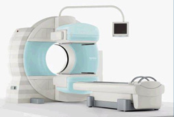
“SPECT-CT nuclear medicine elegantly combines the anatomic accuracy of CT scan with the functional information of nuclear medicine. This fusion of two types of imaging is possible using a hybrid scanner (see photo) and clever computer software and technology. “
“This has revolutionised our practice of nuclear medicine as I showed in our 2009 study.”
The combination of two different imaging modalities is synergistic and in this context
1 + 1 = 3
Our fusion bone scans combine a low dose multislice CT with a regional or whole body bone scan which gives us an excellent tool to help assess the source of a patient’s pain. In my experience SPECT-CT bone scans are the best imaging modality to confirm or localize symptoms arising from
1. Degenerative disease of the spine where the pain is NOT nerve root –it can accurately assess the site of disc degeneration and facet joint arthropathy
2. Fractures at any site but especially stress and fatigue fractures
3. Bone or joint pathology in the hands and feet
4. Lateral hip pain syndromes –it can confirm gluteal tendon attachment problems, hip joint pain, and stress fractures. It will help differentiate pain referred from SIJ, lumbar spine, and regional hip pathology.
Unlike other modalities nuclear medicine abnormalities strongly correlate with a patient’s symptoms. Degenerative soft tissue and joint pathology is universally found in scans of anyone over 40 years but the presence of an abnormality on U/S, CT, Xray, or MRI does not necessarily indicate it is symptomatic. On the other hand a hot spot on a bone scan is usually symptomatic.
SPECT-CT (fusion) nuclear medicine is both more sensitive and more specific than planar nuclear scans for a many clinical problems:
1. Assessment of sclerotic foci demonstrated on x-ray or CT.
2. Differentiation between benign and malignant disease.
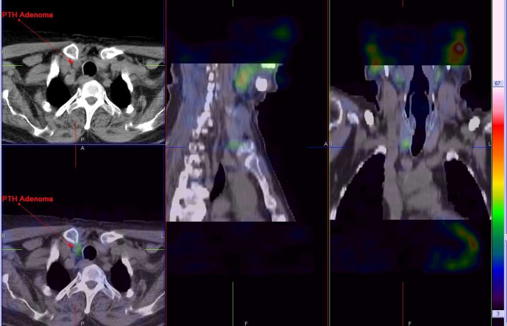
3. Parathyroid adenoma localisation (see images above)
4. Infection Imaging – Gallium and white cell scans
5. Tumour Imaging –MAA and Octreotide scans
6. Liver lesion imaging -Red cell Scans for Haemangiomas in the Liver
7. Brain scans –combined CT and neurolite for vascular dementia
For a more visual tour through SPECT-CT in modern nuclear medicine see this slide show: The New Nuclear Medicine of 2012 .


