
WHAT IS IT?

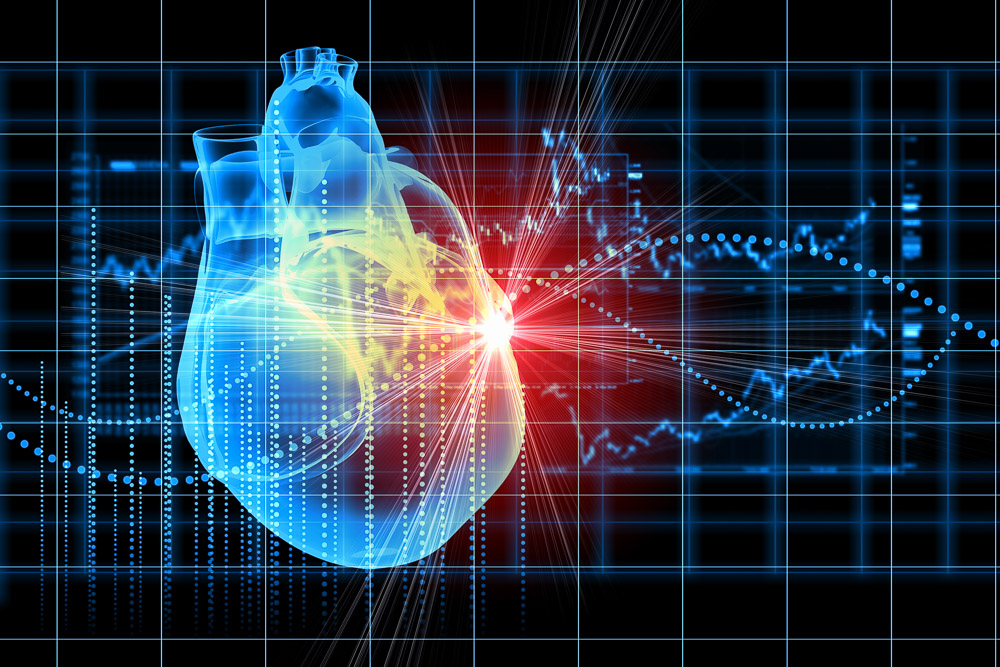
Sestamibi uptake in the left ventricle
A myocardial perfusion scan is a test to evaluate blood flow and contraction of your heart muscle. A radioactive material called a radio-tracer is injected through a vein into your bloodstream. The radio-tracer then locates in your heart muscle in proportion to the amount of coronary blood flow. The radio-tracer produces gamma rays that a gamma camera detects and uses to produce images. Myocardial perfusion scanning has good track record and is a safe way of evaluating your heart. For cardiac testing we use the following radio-tracers: thallium-201, technetium-99m sestamibi (Cardiolite®), and technetium-99m tetrofosmin (Myoview™). These radio-tracers expose the body to a very small amount of radiation. No long-term effects have been reported from these doses. We use these tracer as they are taken up into the myocardium (or heart muscle) as you can see in the image.
For a myocardial perfusion scan we take two sets of images (two separate scans), one while the heart is more active (stress images) and one when the heart is resting. The rest scan is done first and after the “stress” test a second scan is done. The stress images are taken after we either give you a drug or exercise you to increase coronary blood flow and often this is associated with an increase in heart rate. We may ask you to exercise lightly or ride an exercise bike while you are receiving the drug (usually persantin or adenosine) as this help reduce blood flow to your stomach and liver which can interefere with our images. You may develop a flushing, pounding head, dizziness or chest tightness while receiving the drug. We reverse the effect of the drug after this part of the test.
BEFORE THE TEST
We need to know about your overall health, including health problems such as asthma, chronic obstructive pulmonary disease (COPD), diabetes, and kidney disease. Let us know about any medicines you take as some medicines and supplements can cause problems when used with the drugs we use to increase your heart rate during the stress test. You must not drink coffee or tea for 12 hours prior to the test as the caffeine counteracts the drugs we use in the study. We will ask you to fast for 4-6 hours before the test. If your test in in the afternoon then a light breakfast is recommended. If your test is in the morning you should only have a glass of water.
Wear comfortable walking shoes and loose-fitting clothes for the test. You can bring snack with you or we will provide you with a sandwich which can be eaten after the stress part of the test and before the second scan -this will help you recover from the fast/drug/and exercise and improves or images.

This is the gamma camera we use for myocardial scanning
The gamma camera is shown here and you will lie on a bed which will pass through the scanner and the scanner will move around the left side of your chest to take the cardiac images. Sometimes, the room feels cold because of the air conditioning needed to cool the machines.
DURING THE TEST
The procedure steps through these stages:
- The placement of an iv line (so we can give all the injections through a small cannula)
- An injection of radio-tracer
- A scan of your heart (15-20 minutes)
- An iv infusion of a drug (the doctor will do this) while you are being monitored on an ECG machine
- You may be asked to pedal on an exercise bike or walk on a treadmill for several minutes. This is not strenuous unless we are doing an exercise test without using a drug (persantin).
- A further injection of tracer and reversal of the cardiac drug
- A short break -a snack or drink of water can be taken at this point
- A second scan of your heart (15-20 minutes)
- Occasionally extra views are needed (further 10-15 minutes)
The whole process usually takes 2 to 3 hours –allow a full morning or afternoon.
AFTER THE TEST
Your doctor who referred you for the scan will receive the results the same or following day via email. He/she can access the images via the web using an online-portal. You can pick up a hardcopy (disc and report) if required.
WHAT DOES IT MEAN?
 This scan assesses cardiac muscle blood flow and does not show detailed coronary anatomy. Information this scan can provide is usually enough to answer one or more of the following questions.
- Is my chest pain due to a restriction in coronary artery blood flow?
- How much restriction of blood flow is my coronary artery disease causing?
- How well are my grafted or stented coronary vessels working?
- How well is my heart contracting /functioning?
- Is there any cardiac risk if I am going to have major surgery?
- Is my cardiac medication working well enough?
FEEDBACK
Let us know about how your experience could be improved or give us some feedback at info@driainduncan.com.au.
Myocardial Perfusion SPECT Scan Information for Patients can be downloaded as a pdf file. See DOWNLOADS.