When I started in nuclear medicine it was often cynically stated by other physicians that nuclear medicine was “unclear” medicine. Unfortunately like so many jokes this one had more truth than I was comfortable with. Nuclear medicine was great at finding the problem but not telling you what it was. We could find “hot spots” on lots of scans but it was up to us to best guess what the “hot spot” might be. Of course like most doctors we fancied we were pretty good at best guessing but in retrospect we were not as good as we thought -my research on SPECT-CT in nuclear medicine showed we were wrong up to 25% of the time. Others have similar findings.
The good news is this era has passed. Why it took so long remains somewhat of a mystery. CT scans (produced by xrays) and nuclear medicine scans (produced by gamma rays) provide complimentary information.
Combing the two types of scanners seems obvious but it is only with the advent of modern computer technology, clever software and the falling price of all the sophisticated components that has made this possible for every day use.
The advent of IMAGE FUSION means that we can see exactly where and usually what the “hot spot” or “cold spot” is. Gradually we are discovering new benefits and improving the effectiveness of nuclear medicine. Inside nuclear medicine this is a quiet revolution and for those on the outside it is a revelation.
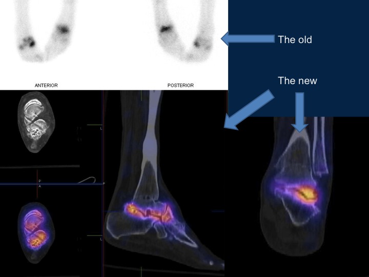
Two examples of the “old” and the “new” showing the power of using better scanning techniques using SPECT-CT. The “old” planar images are shown in black and white and the “new” SPECT-CT fusion images show CT (xrays) in black and white and nuclear scans (gamma rays) in colour. Hundreds of slices in all planes can be generated using SPECT-CT. The mix of SPECT and CT fusion can be dialled up or down. The amount of information generated is hundreds of times larger than with planar scans which are more like a photograph.
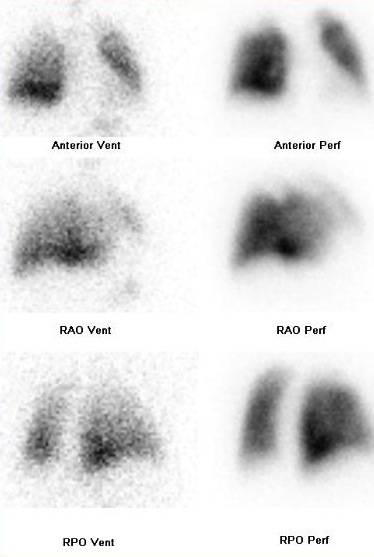
Both cases represent scans done in the SAME patient. At the top is a bone scan of a painful left hindfoot with the old and new shown together. The second image is a conventional lung scan showing both perfusion (blood flow) and ventilation (air flow) scans.
The third is the same patient as the second but with a representative slice through the lung apices. There are two planes shown with one each of perfusion and ventilation. The cold area on the perfusion scan represent a blood clot or pulmonary embolism. See if you can see the abnormality in both the old and the new scans.
For doctors interested in the changing clinical use of lung scans see also NEWS.


