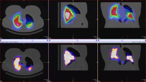
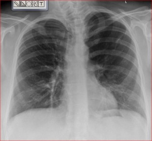
This middle aged female was referred with shortness of breath and a CXR reported as showing oligaema in the left upper zones that could indicate pulmonary embolism. The reconstructed planar lung scan showed a large matched defect corresponding to the left upper lobe. The SPECT-CT scan shows a large airspace is responsible for the abnormality -most likely a (post-infectious) pneumatocoele or large bulla.
- Lung scan with matching LUZ defect
The planar images would be reported as indeterminate but the SPECT-CT demonstrates the specific cause and excludes pulmonary embolism.


