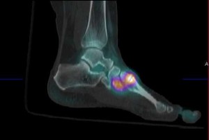
The Clinical Impact of SPECT scanning in Nuclear Medicine Practice.
Iain Duncan & Barry Flynn, Canberra , ACT, Australia, 2009.
Objective:To prospectively evaluate the clinical value of single photon emission tomography-CT nuclear scanning (SPECT-CT) in private nuclear medicine practice.
Methods: All (consecutive) patients attending a single practice between August and November 2009 were included. The reporting clinician made a diagnosis and provisional report based on standard planar imaging and a decision whether to proceed to SPECT-CT (using a Siemens Symbia T2) was then made. After the SPECT-CT a final report was issued and a simple questionnaire completed. Four questions were asked:
1) Has the SPECT-CT altered the primary (planar) diagnosis?
2) Has the SPECT-CT significantly altered the interpretation of the scan?
3) How many extra lesions are seen in the SPECT-CT compared to the planar images?
4) Why was SPECT-CT not done in this patient? The reporting clinician’s answers were recorded and later analysed.
Results: 1007 patients were included. No patients were excluded from the study. 62% were bone scans and 38% others. 71% of bone scans and 21% other scans underwent SPECT-CT (38% of others excluding cardiac scans). In patients undergoing
SPECT-CT this changed the final diagnosis in 25% of bone scans and 11% in all other scans. SPECT-CT significantly altered the interpretation of the scan in 66% of bone scan and 61% of others. On average SPECT-CT identified an additional 0.9 lesions in bone scans and 0.2 lesions in other scans. There were slight differences between two reporting physicians.
Conclusions: We concluded that SPECT-CT has had a major clinical impact in our practice of nuclear medicine.
This is a summary of a paper presented at the Australasian Society of Nuclear Medicine Conference in Auckland, New Zealand in April 2010.

