MAY 2013 to DECEMBER 2015 NEWS ARCHIVE
November & December 2015
Most of us are time poor and thinking on this recently I came up with a concept of 10 minute updates for our referrers. You don’t need an exhaustive lecture to get your head around something. We have now progressed the concept to reality and hopefully these short presentations can update referring practitioners on medical imaging from the basics to the most recent technologies, scientific advances, and concepts. It is also an opportunity for forging a better relationship between users and providers of medical imaging, something that is definitely needed. In a little more than half an hour 3 subjects can be covered and there is still plenty of time for informal discussion and chit chat. Kevin Osborn and I launched this on the 2nd December at Garran Medical Imaging. The audience was small (6 including Kevin and I) but you have to start somewhere. After a break in January 2016 we plan to recommence in February, once a month. We could even come to you either in the real or virtual -all requests considered! Just let us know what you would like to hear about (info@driainduncan.com.au). My favorites are, of course, recent advances such as elastography, xSPECT nuclear imaging, PRP therapy and as always any musculoskeletal ultrasound topic.
If you managed to read this sentence I congratulate you and wish you the very best wishes of the season.
October 2015
I have started offering patient’s ultrasound guided platelet rich plasma (PRP) injections at Garran Medical Imaging using a new system from regenlab which fulfils the criteria I have outlined in my update this month. I am confident that this can have a clinical impact where alternatives are extremely limited (steroids, polidocanol, hyaluronate, glucose, autologous blood).
And what about that win against Wales at the World Cup.

Wallabies psyched up for a momentous battle v Wales
September 2015
Some new website additions: New abstract on patellofemoral pain. I was able to attend both the ASUM ultrasound meeting in Sydney and the following week the ASA Special Interest Group meeting, where I gave two workshops, helped in another, and participated in a plenary forum. For those who requested a closer look at some of the images for ankle and foot you can find them here.
For those interested in pulmonary embolism I discovered this useful reference for the diagnosis: the PERC rule to exclude PE if no criteria are present and pre-test probability is <15%.
August 2015
August has been a lot of hard work but great fun. Garran Medical Imaging had a late start in mid July but is now up and running. Plenty of minor troubles but the staff have been amazing and the future looks very bright. There are lots of first for the business which has both embraced principles of customer service from other industries and endeavoured to put the patient at the centre. Needless to say Garran Medical Imaging will be looking at new ways to use cutting edge technology to deliver clinical benefits for patients and doctors. This is my “raison d’être.” Two such technologies are xSPECT bone imaging and shearwave elastography which are feature stories on the website this month. These technologies are currently available at only a few locations in Australia. While xSPECT will raise the bar of nuclear bone imaging, shearwave elastography provides a new way of assessing the liver for those with chronic hepatitis and liver disease.
July 2015
I have teamed up with Dr Kevin Osborn and an enthusiastic team at a new imaging centre of excellence. Garran Medical Imaging is located at Garran Shops (near The Canberra and National Capital Private Hospitals). Garran Medical Imaging aims to be part of the solution for doctors and patients. Kevin and I would like to better partner with referring doctors to help solve clinical problems and improve/de-stress a patient’s journey through the medical system.The practice has been designed to provide excellence at every interaction and from all angles. The patient journey from referral through booking, check-in, scanning, completion and delivery of results has been carefully mapped to ensure a comfortable and stress free experience. We are aiming to make the diagnostic quality of our scans world class.

June 2015
I have commenced a new “ultrasound blog” for the benefit of all the sonographers I work with and those that I don’t. Feel free to send me feedback via email. You can find it under “Sonography grabs” in the categories search menu or via the “Information for Sonographers and Sonologists” section in Resources and Updates.
Garran Medical Imaging is now in the final stages of gestation with equipment partly installed. I am so excited and at the same time terrified of some oversight or further delay (we are only going to open 4 months late). The website will launch this week and our PR campaign is under way. I cannot believe that a medical imaging practice could have had more love and attention prior to debut. Lets hope the momentum rolls on…
May 2015
In conjunction with Dr Kevin Osborn I am proud to announce the creation of Garran Medical Imaging which is a new Canberra Medical Imaging practice designed to deliver best possible service and diagnosis using advanced imaging technology. Together with Nick Ingold and an amazing team we aim to deliver cutting edge diagnostic imaging in a more caring and supportive way. Let us show you why we love our jobs and how we can help make your diagnostic journey the best one possible. Planning has been going on for more than a year and final building construction will be complete in June. The best possible equipment along with truly dedicated staff will open the doors of Garran Medical Imaging for business in July 2015. The website of Garran Medical Imaging will launch soon.
April 2015
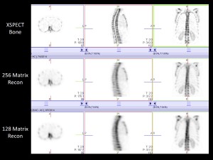
I attended the ANZSNM (The Australian and New Zealand Society of Nuclear Medicine) annual Scientific meeting in Brisbane between the 17th and 20th April. As well as getting my dose of educational updates I listened to those who have had the first experience with X-SPECT technology in Australia. This is particularly exciting as Garran Medical Imaging (GMI) will be joining the early adopters of this awesome technology. This aligns with the mantra of GMI which is all about exploring new ways of using medical imaging technology to improve clinical outcome for both doctors and patients. We want to be part of the solution. GMI will be introducing several new technologies into clinical practice and while the technology has been scientifically validated no one is certain how it is best applied or how much clinical difference it will make. I will be encouraging a dedicated team of professionals to collect information that helps deliver solutions. In the meantime check out these images below.

XSPECT BONE v XSPECT v SPECT
The XSPECT bone images on the top row compare with the best current systems on the bottom row. The middle row is the SPECT images on the new system without the X-SPECT processing. These images are prior to fusing with the CT part of the scan. I am sure you can see the difference (click on the image to enlarge).
On another note entirely a new study has shown stretching is unproven in preventing tendon injuries but that footwear is important.
March 2015
Exciting news. A brand new medical imaging practice will be opening in Canberra later this year. This practice has been planned from the ground up in every respect: custom-built modern premises, state of the art equipment and an inclusive team environment. Garran Medical Imaging will have a small and dedicated team of professionals who will set a new benchmark for the industry. Needless to say Iain will be keen to share news and detail over the next few months as the jigsaw comes together. For those professionals who would like to participate a select number of jobs have been advertised here.
February 2015
The website has had a makeover in January -the organisation is different to cope with more content and there are lots of new illustrations. New additions include an abstract about breast tomosynthesis which will hopefully improve the journey for women requiring investigation and screening for breast cancer. This is not yet widely available but will come on stream as radiology centres update their equipment over the next few years.
January 2015
Quiet start to the new year and now accelerating with lots of changes to the website, both in content and organisation. The New Year is also a chance for lots of reading both for technical and spiritual growth. James Alutcher captures some great LIFE TIPS to help us reinvent ourselves in the new year.
Back pain is such a common problem and to clarify the role of bone scanning and for a quick update check out my latest post.
I have also written a paper on scanning of prosthetic joints (hips, knees, and some back surgeries) with a quick reference flowchart. Unfortunately there are no gold standards for sorting out complications of joint replacement and post-op pain. Nuclear medicine is the next stop after a plain x-ray. See here.
I get a lot of questions about terminology and classification. We all end up using terminology well known to our individual craft groups but not always well understood by others. To tackle this I will progressively add to the website glossaries (found in either the referring doctors area or the Search by category MENU) my commonly used reporting classifications and terms I use, for example stress fractures.
[…]
READ MORE


